
3D Printed Vaginal Rings Could Treat Bacterial Infections


There are plenty of examples in which 3D printing has been used to develop drug delivery systems, but this research out of Hungary is tackling the issue from a new direction. We’ve seen 3D printed spermbots used to deliver cervical cancer-fighting drugs. Now researchers from the University of Debrecen and University of Szeged are using fused deposition modeling (FDM) 3D printing to make a system that delivers drugs vaginally. In their paper, the team explains that vaginal drug delivery systems can offer a “long-term and constant liberation of the active pharmaceutical ingredient even for months.”
“For our experiment, FDM 3D printing was used to manufacture the vaginal ring samples from thermoplastic polyurethane filament, which enables fast manufacturing of complex, personalized medications. 3D printing can be an excellent alternative instead of industrial manufacturing, which is complicated and time-consuming,” they wrote in the abstract.
Vaginal infections, while not typically deadly, are widespread and often last a long time. They can also be hard to cure, uncomfortable, and anxiety-inducing for the patient, no doubt. Additionally, because of the vagina’s anatomy and “the bacterial diffusion between the rectum and the vagina,” relapses of infections can often occur, and the researchers say that 30% of women treated with antimicrobials, like clindamycin and metronidazole, will experience a recurrence of the vaginal infection for various reasons.
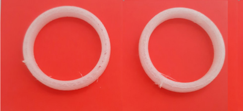
Figure 1: Image of the 5 mm tall printed samples.
As the team explains, the first patent for a vaginal ring drug delivery system, in which users can “initiate or discontinue use whenever desired,” was given in 1970. Unfortunately, design and manufacturing methods are different for each product, as medical agencies haven’t offered relevant regulations or guidelines. While injection molding and hot-melt extrusion have been used to fabricate these rings, the researchers went with fused deposition modeling, or FDM, 3D printing, which is a popular AM technology and “enables the manufacturing of oral dosage forms, intrauterine systems, implants or vaginal rings.”
“For the manufacturing of a drug delivery system with 3D printing, researchers incorporate the API into the filament with hot-melt extrusion, and then the API-containing filament is printed with FDM technology,” the researchers explained. “The high extrusion temperature used in FDM (more than 120 ◦C) and in hot-melt extrusion (more than 150 ◦C) enables only heatstable APIs incorporation into the polymeric filament. Polymer modification or different added excipients can also decrease the printing temperature, but the lowest used printing temperature was 165 ◦C, which is still not adequate for a lot of API’s. Even though many research projects are trying to solve the problem of high printing temperature, the most promising solution would be to manufacture a carrier system in which all types of APIs can be incorporated.”
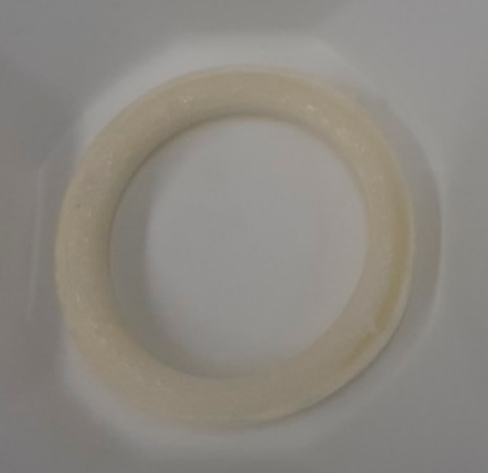
Figure 2: Image of the manufactured samples.
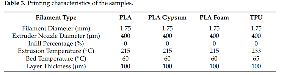
These researchers decided instead to 3D print vaginal rings to act as drug carrier systems for bacterial vaginosis, and fill the pre-printed samples with jellified APIs (vaginal gels), specifically the antibiotics metronidazole and chloramphenicol (CAP), as the benefits of vaginal gels include comfort, ease of fabrication, and “the ability to cover the surface of mucosal membrane.” Digital models for the rings—based on the commercially available NuvaRing but with chamfered edges—were designed in SOLIDWORKS, and the team printed batches of four drug delivery systems out of polylactic acid (PLA), PLA Gypsum, and PLA Foam from Philament, and thermoplastic polyurethane (TPU) 80A LF from Ultrafuse FFF on a Craftbot 3 system.
They prepared four different formulations of the APIs:
- Carbopol 934 and a 1 w/w% solution, manufactured with distilled water at 500 rpm shaking
- 3 w/w% chitosan and 4 w/w% hydroxyethyl cellulose (HEC)
- 1 w/w% chitosan and 4 w/w% HEC, with distilled water, 300 rpm shaking, and manual mixing
- 1 w/w% agar-agar solution, prepared with distilled water and heating up to 100°C
Upper and lower parts of the vaginal ring were pre-printed, and 5 g of chloramphenicol or metronidazole were measured out before being suspended with 20 g of gel. Then, the various suspensions were put into syringes, and 1.5 g of each gel was added to the lower parts of the rings, before the upper parts were added and closed manually.
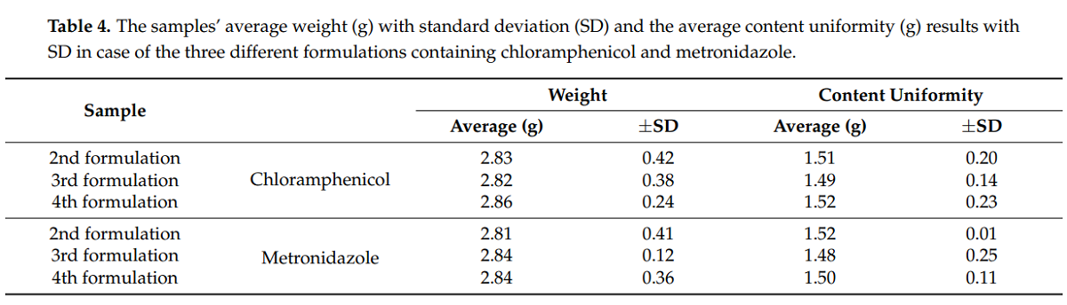
The researchers individually weighed ten samples from each formulation, before they were “opened for content uniformity determination.” Then they carried out thermogravimetric (TG) and heatflow (DSC) analysis on the samples, which certified the “need for manual filling,” before the samples were analyzed by a dissolution apparatus; all were “considered non-similar based on the pairwise comparison.” Prolonged MTT assay on HeLa cells was used to determine the biocompatibility properties of the samples, which were found to be non-toxic. The team also performed contact angle measurements with the sessile drop method, and measured the results with a microCT system.
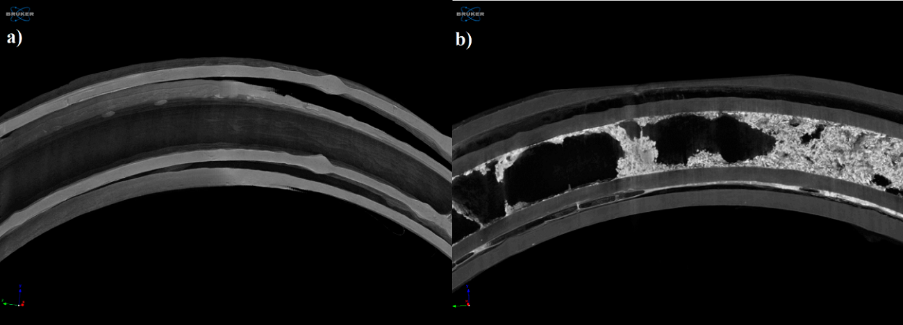
Figure 7: Reconstructed microCT image from a vertical cut of the printed sample (a) and examined sample after the dissolution test (b). Image pixel size is 5 µm.
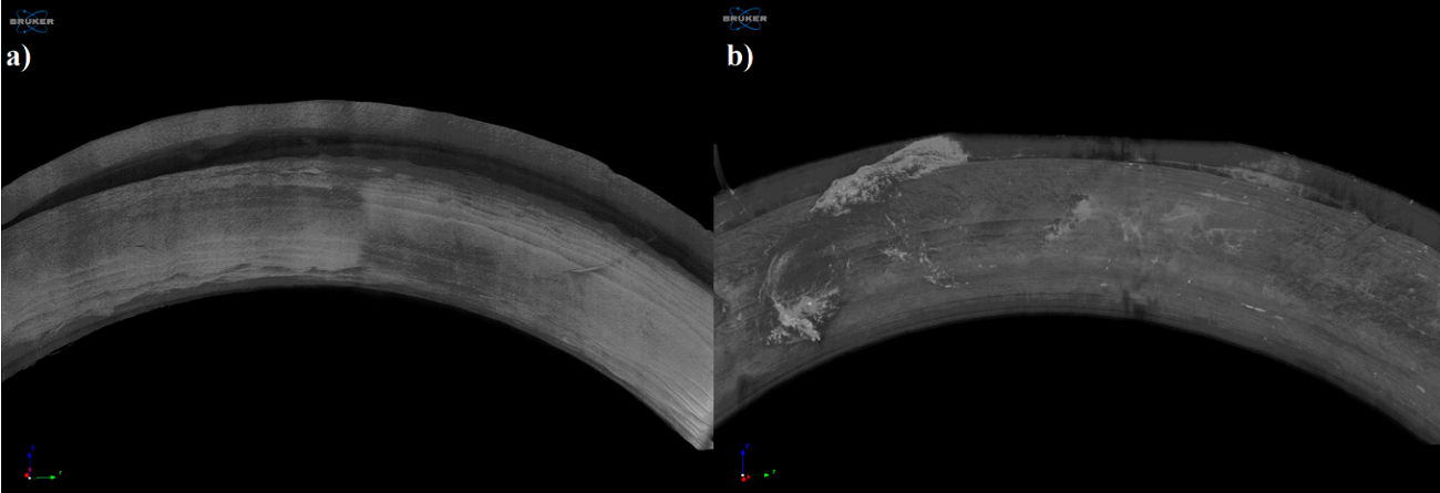
Figure 8: Reconstructed microCT image from the upper surface of the printed sample (a) and examined sample after the dissolution test (b). Image pixel size is 5 µm.
In vitro dissolution testing was also performed, and UV spectrophotometry was used to determine absorbance of the release drug. In Figure 7, you can see localization of the remaining gel after dissolution, but on the surface of the dissolved sample in Figure 8, no change was detected.
The team also evaluated the microbiology of the samples in the 3D printed rings, in order to “examine the effect of vaginal rings containing different compounds,” and found that E.coli majorly decreased “in the presence of three tested rings.”
“The tested samples were as follows: (a) empty vaginal ring; (b) metronidazole containing vaginal ring; (c) 3 w/w% chitosan and 4 w/w% hydroxyethyl cellulose-containing vaginal ring; (d) 3 w/w% chitosan and metronidazole containing vaginal ring.”
The team determined that TPU was better than PLA for 3D printing the rings, because it was more flexible, and that “the 32nd printing parameter adjustment was acceptable for the manufacturing.” As for the formulations, the first was determined to be too watery, while the second was homogenous and consistent. The third was more watery than the second but less than the first, and after cooling, the fourth became a semisolid gel.
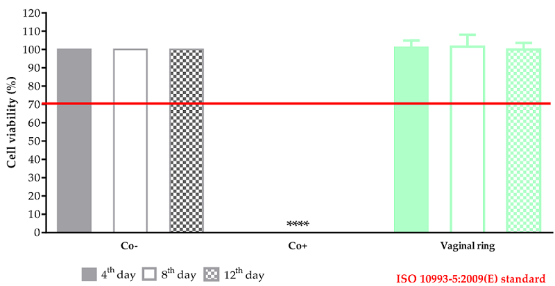
Figure 11. The prolonged cytotoxicity of the 3D printed sample by TPU. MTT cell viability tests were performed on days 4, 8, and 12. Cell viability was expressed as the percentage of untreated control (Co-). As a positive control (Co+), Triton X 100 (10% w/v) was used. Data were expressed as means of four independent experiments ± SD.
Different from the original MTT assay, they also performed a prolonged cell viability test to learn more about the cytocompatibility (property of not being harmful to a cell) of the 3D printed vaginal rings, and found them to be non-cytotoxic, based on the ISO 10993-5:2009(3) standard.
Overall, the team determined that FDM 3D printing could be used to successfully fabricate vaginal rings out of TPU, and suggested that metronidazole was the way to go in terms of filling the drug delivery system.
“The special experimental arrangement ensures that all kinds of API can be utilized during printing without heat damage or API loss, confirmed by TGA results. Based on the dissolution curves, the used API and jellifying agent can modify the dissolved API amount. Based on the MTT assay results, TPU polymer can be considered cytocompatible. The microbiological evaluation confirmed that metronidazole and chitosan have a synergistic effect against E. coli.”
Subscribe to Our Email Newsletter
Stay up-to-date on all the latest news from the 3D printing industry and receive information and offers from third party vendors.
Print Services
Upload your 3D Models and get them printed quickly and efficiently.


You May Also Like
The Drone Economy Needed a Scalable Manufacturing Backbone. ADDMAN Built One
When ADDMAN closed its acquisition of Forecast 3D in January 2026, the headlines focused on fleet size and Southern California footprint. Six months later, those metrics feel almost beside the...
The Drone Industry is Showing Where 3D Printing Delivers Real Value, AM Research Report Finds
The rapid rise of drones is creating one of the biggest opportunities for additive manufacturing (AM). Whether they’re used on battlefields, inspecting bridges or crops, or delivering supplies, drones need...
The Longevity Economy Needs a Factory
Longevity has become one of the biggest stories in healthcare. Every week seems to add a new announcement about an anti-aging therapy, an AI-powered drug discovery platform, or a startup...
The SLS Market: Game of Trucks
This is truly an exciting moment in the SLS market. With HP‘s release of the 1200 and Formlabs‘ release of the X1, we can see the SLS market heating up....


