SLA 3D Printing For Microneedle Transdermal Drug Delivery Systems

In the recently published ‘A 3D printed microfluidic-enabled hollow microneedle architecture for transdermal drug delivery,’ researchers explore innovative methods for delivering medication into the bloodstream via the skin. In this study, they work on the micro-level to create ‘new degrees of freedom’ in delivery.
As medicine continues in the direction of patient-specific treatments, 3D printing continues to play an enormous role—including medical devices, implants, innovations in tissue engineering like scaffolding, and much more. In this study, the researchers continue to refine the use of needles for transdermal use.
Conventionally, they have been fabricated with materials like plastics, metals, ceramics, and more. With the advent of biocompatible polymers, microneedles are being more widely used due to greater disposability, affordability, and the potential for customization—pointing toward patient-specific benefits overall.
Microfluidic devices are behind many of the new capabilities in drug delivery systems, allowing for mixing and transporting of the required small amounts of fluids.
“For example, microfluidic mixing was used to directly synthesize nanoparticles with tunable physicochemical properties such as particle size, homogeneity, and drug loading and release at the point of delivery,” state the researchers. “Additionally, the combination of microneedles and microfluidic mixing is beneficial in areas such as combinational therapy-based subcutaneous/transdermal administration for preclinical testing of biologic treatments.”
New systems are being developed too for ‘codosing,’ allowing for patients to receive several medications at once. Microfluidics and innovative drug delivery systems make the process more affordable, simpler, and less open to error. This study yields microfluidic-enabled microneedle devices printed via single-step stereolithography (SLA) from an ‘elaborate hollow microneedle design,’ resulting in a refined microneedle array.
“This architecture allows the modulation of the input fluid solutions’ flow rates to facilitate programmable drug delivery in future combinational therapy-based applications,” stated the researchers.
While there are benefits to SLA 3D printing, the research team was tasked to refine the process further for this study, creating a new microneedle design and print set up.
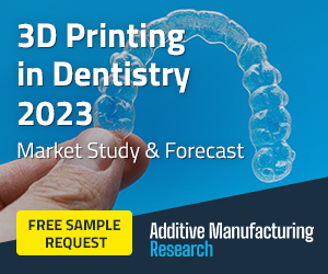
3D-printing of microfluidic-enabled hollow microneedle devices. (a) CAD model of a representative microfluidic-enabled microneedle device as an input to the SLA printer. (b) The printed device with three microfluidic inlets converging into a 3D spiral chamber and to a hollow microneedle array outlet. (c) Close-up of the inlet junction visualizing the convergence of red-dyed, clear, and blue-dyed solution streams. (d) Close-up of the hollow microneedle array.
The research team was able to create up to 12 devices (with dimensions of 1.5 × 1.2 × 3.1 cm) using class IIa biocompatible resin in a single print, in 2.5 hours.
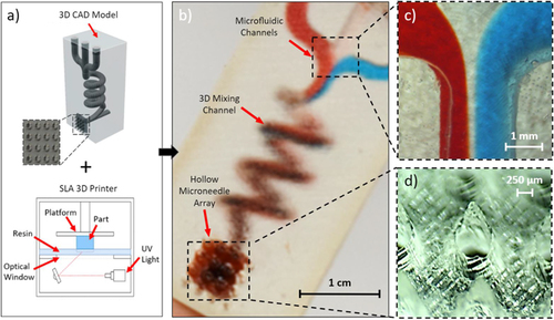
Characterization of 3D-printed hollow microneedle arrays. Images of sheared-cylinder microneedles printed at (a) 0°, (b) −45°, (c) +45°, and (d) 90° angles with outlined profiles (insets show the corresponding print setups). SEM images of the (e) conical, (f) pyramidal, and (g) basic syringe-shaped needle arrays. (h) Average needle heights for each design (for a subset of 25 microneedles per array). (i) CAD model of the syringe-shaped design and the SEM image of the tip (∼50 μm radius of curvature). (j) CAD model of the fine-tip syringe-shaped design (additional features highlighted) and the SEM image of the tip (∼25 μm radius of curvature). (k) SEM image of the fine-tip microneedle array. (l) Average microneedle heights across three separate fine-tip microneedle arrays (for a subset of 25 microneedles per array). Error bars indicate ±standard deviation.
Scanning microscopy displayed success in both design and 3D printing of the arrays.
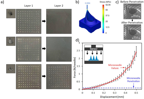
3D-printed microneedle mechanical characterization: penetration and failure. (a) Penetration test of the pyramidal, conical, and fine-tip syringe-shaped microneedle arrays across two layers of the parafilm with 5 N of applied force. (b) Mechanical simulation of the fine-tip syringe-shaped microneedle, visualizing the occurrence of maximal stress at the tip. (c) SEM image of a microneedle before and after penetration testing (demonstrating no tip failure). (d) Axial force vs displacement curve for a 3 × 3 array of syringe-shaped microneedles. The failure point and penetration force are noted. The inset image illustrates the compression test setup.
“Penetration and fracture tests confirmed the microneedles’ mechanical robustness for practical application. An example microfluidic-enabled microneedle device was printed with our devised scheme that facilitates homogeneous mixing of multiple fluids under different flow rates, followed by transdermal delivery of the mixed solution. Comparisons of various flow rate ratios with colored dye solutions showed tunable control over the relative concentrations of solutes delivered. Ex vivo confocal laser scanning microscopy of three fluorochrome model-drug solutions on porcine skin further validated the platform’s ability for transdermal drug modulation and delivery,” concluded the researchers.
“This 3D printed device is particularly applicable to preclinical investigations centered on combinational drug therapy, where the in-situ combination of multiple drugs and the tuning of their physicochemical properties lead to more effective outcomes than single or premixed agents alone. For example, the controlled multifluidic synthesis of nanoparticles can tune the release mechanisms of various drugs for wound healing applications.”
What do you think of this news? Let us know your thoughts! Join the discussion of this and other 3D printing topics at 3DPrintBoard.com.
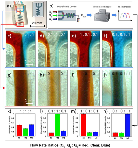
Mixing characterization of the 3D-printed microfluidic architecture. (a) Photograph of the SLA-printed microfluidic mixing architecture with CAD model in the inset. (b) Schematic of the solution concentration quantification method. (c)–(f) Microscopic images of the 3D spiral chamber’s inlet junction under various flow rate ratios of red-dyed, clear, and blue-dyed solutions (Q1:Q2:Q3). (g)–(j) Microscopic images of the 3D spiral chamber’s outlet under the corresponding flow rate ratios. (k)–(n) Normalized fluorescence (FL) intensities of rhodamine B (RB), fluorescein isothiocyanate (FITC), and methylene blue (MB) present in the solutions obtained from the outlet. Error bars indicate ±standard deviation (n = 3).
Subscribe to Our Email Newsletter
Stay up-to-date on all the latest news from the 3D printing industry and receive information and offers from third party vendors.
You May Also Like
3D Printing News Briefs, April 13, 2024: Robotics, Orthotics, & Hypersonics
In 3D Printing News Briefs today, we’re focusing first on robotics, as Carnegie Mellon University’s new Robotics Innovation Center will house several community outreach programs, and Ugogo3D is now working...
Rail Giant Alstom Saves $15M with 3D Printing Automation Software 3D Spark
3D Spark has entered into a three-year deal with the rail giant Alstom. Alstom, a transport behemoth with annual revenues of $16 billion, specializes in the manufacture of trains, trams,...
Meltio Expands Global Reach with New Partnerships in the Americas and Europe
Spanish 3D printing manufacturer Meltio has expanded its sales network across the globe. With the addition of three new partners in the United States, Brazil, Argentina, and Italy, Meltio aims...
3D Printing Webinar and Event Roundup: April 7, 2024
Webinars and events in the 3D printing industry are picking back up this week! Sea-Air-Space is coming to Maryland, and SAE International is sponsoring a 3D Systems webinar about 3D...