

Osaka University: Vascularized Cardiac Construction with LbL & 3D Printing
Authors Yoshinari Tsukamoto, Takami Akagi, and Mitsuru Akashi, all from Osaka University, experiment with bioprinting in cardiac medicine, explaining their findings in the recently published ‘Vascularized cardiac tissue construction with orientation by layer-by-layer method and 3D printer.’
As tissue engineering continues to evolve in labs around the world, reaching the goal of 3D printing human organs hovers ever closer; and while such progress may seem just out of reach for many scientists, the fabrication of 3D tissue in new studies continues at a rapid pace. In this research, the authors continue where they left off in previous work, forging ahead to further refine cardiac tissue engineering.
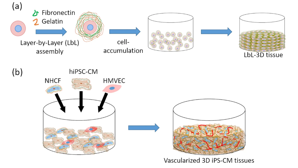
Schematic illustration of the fabrication process for layer-by-layer (LbL) 3D tissue using fibronectin (FN) and gelatin (G) coating technique and cell accumulation technique. (b) Schematic illustration of fabrication of 3D cardiac tissue with a blood capillary network using LbL coated cells and cell accumulation technique.
Bioprinting cardiac tissue with a heart specific structure, cell orientation, and a vascular network, the authors used layer-by-layer fabrication (LbL), cell accumulation, and 3D printing. A hydroxybutyl chitosan (HBC) gel frame was created via 3D printing to control the orientation of the cells ‘linearly.’
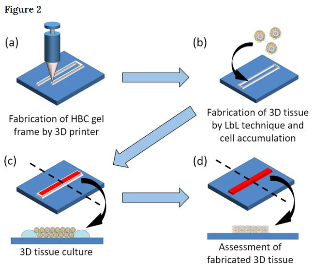
Schematic illustration of fabrication of orientation-controlled 3D cardiac tissue using 3D printing technology. (a) 3D printing of HBC using a robotic dispensing 3D printer. (b) Fabrication of 3D multilayer tissue using LbL coated cells and cell accumulation technique. (c) Cultivation of orientation-controlled 3D tissue. (d) Assessment of shape and contractile properties using a histological technique and image processing.
“HBC has the ability of sol-gel transition depending on the temperature,” stated the authors.
The use of HBC gel was particularly interesting as the researchers used a robotic dispensing printer, cooling ink to 4 °C with a Peltier element. Evaluation by the authors showed that line width of the ink was around 1mm, with the potential for lamination of up to eight layers.
“A ninth layer could not be laminated because the HBC gel wall melted. The reason for this is that the ninth layer is far from the substrate and melts because it cannot receive temperature control,” explained the researchers. “From our previous studies, however, the thickness of 3D tissue is limited to 100 μm. For this reason, the 3D modeling ability of HBC gel is sufficient to fabricate 3D tissue using an LbL technique and cell accumulation technique.”
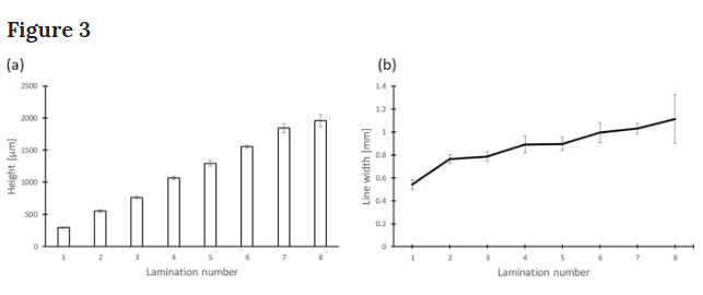
Observation and analysis of a laminated 5% HBC gel wall printed by a robotic dispensing 3D printer. (a) Height of laminated HBC gel wall observed from the horizontal direction. (b) Line width of laminated HBC gel wall observed from the vertical direction.
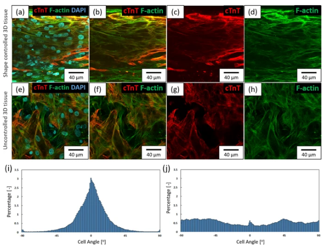
Shape controlled 3D cardiac tissue image stained with fluorescent labeling phalloidin and anti-cardiac troponin T (cTnT) antibody obtained from confocal laser scanning microscopy (CLSM). (a–d) Shape controlled 3D cardiac tissue using a 2 × 15 mm HBC gel frame. (e–h) Uncontrolled 3D cardiac tissue. (a,e) The merged images of F-actin, cTnT and DAPI. (b,f) The merged images of F-actin and cTnT. (c,g) The cTnT images. (d,h) The F-actin images. (i,j) The graphs of the local alignment angles of F-actin fibers in shape controlled 3D cardiac tissue are shown underneath the CLSM image by image analysis.
Next, the researchers created a vascular network for their 3D printed cardiac tissue, adding hiPSC-CMs and NHCF coated FN-G nanofilms co-cultured with HMVEC in a 1.5 × 15 mm rectangular HBC gel frame (5%). Employing a 1.5 mm short side rectangular HBC gel frame, the researchers were able to control 3D cardiac tissue.
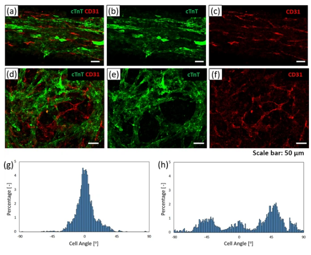
Shape controlled 3D cardiac tissue with vascular network image stained anti-cardiac troponin T (cTnT) antibody and anti-CD31 antibody obtained from LSCM. (a–c) Shape controlled 3D cardiac tissue using a 1.5 × 15 mm HBC gel frame. (d,e,f) Uncontrolled 3D cardiac tissue. (a,d) The merged image of cTnT (green) and CD31 (red). (b,e) The cTnT image. (c,f) The CD31 image. (g,h) The graphs of the local alignment angles of vascular network (CD31) in shape controlled 3D cardiac tissue are shown underneath the CLSM image by image analysis. (g) The graph of orientation-controlled tissue. (h) The graph of uncontrolled tissue.
“From the result of CD31 stained images, vascular network formed in both tissues. In the case of orientation-controlled tissue, the vascular network has an oriented structure similar to cardiomyocytes according to image analysis,” concluded the authors. “In the case of uncontrolled tissue, on the other hand, the vascular network does not have an oriented structure.
“This 3D cardiac tissue has the potential for usage in transplantation medical care and drug assessment because it has the native heart organ-like structure and vascular network for the fabrication of thicker and larger 3D tissue. Therefore, we believe that the 3D cardiac tissue with orientation and vascular network would be a useful tool for regenerative medicine and pharmaceutical applications.”
3D printing of cardiac tissue has been the focus of other research projects, from phantoms used by surgeons to patches and cellularized hearts, regenerated muscle tissue, and much more. What do you think of this news? Let us know your thoughts! Join the discussion of this and other 3D printing topics at 3DPrintBoard.com.
[Source / Images: ‘Vascularized cardiac tissue construction with orientation by layer-by-layer method and 3D printer’]Subscribe to Our Email Newsletter
Stay up-to-date on all the latest news from the 3D printing industry and receive information and offers from third party vendors.
Print Services


You May Also Like
New Business: Temporary, Migratory, & Modular 3D Printed Architecture
If we look at potentially emerging 3D printing businesses, then architecture has not been fully explored. Yes, there is a lot of house 3D printing going on worldwide. From deployable...
3D Printing News Briefs, April 19, 2025: Material Extrusion Standard, Metal Powder, & More
In today’s 3D Printing News Briefs, we’re covering a proposed standard for material extrusion, before moving on to business and metal powder. We’ll end with a commercial store’s robotic 3D...
Japan Unveils World’s First 3D Printed Train Station
Japan is now home to what we believe is the world’s first train station built with 3D printing technology. Located in Arida City, just south of Osaka, the new Hatsushima...
restor3d Raises $38M to Expand 3D Printed Orthopedic Implants
Backed by $38 million in new funding, restor3d is pushing ahead with the launch of four personalized implant lines, set to roll out in 2025 and 2026. This latest venture...