Researchers Study the Effectiveness of 3D Printing in Cardiac Phantoms

 The heart wants what the heart wants, as they say, but in many cases, it may also want some help to continue performing its natural duties in pumping blood throughout the body and removing carbon dioxide. The many miracles of modern science allow medical professionals and surgeons to save patients by repairing issues with the heart, and the tools they use to do so continue to progress with technology like advanced imagery, 3D printing—and sometimes the two together.
In ‘Optimisation of CT protocols for cardiac imaging using three-dimensional printing technology,’ by Kamarul Amin Abdullah of the University of Sydney, a three-tiered study was performed, beginning with 3D printing a cardiac phantom. Next, it was placed within the Lungman phantom and scanned, allowing researchers to reconstruct data and then measure and compare to figure out dose reduction potential. Last, the use of algorithms with varying strengths and ‘low-tube voltage for dose optimization studies’ was evaluated.
Phantoms are commonly used to refine imagery such as CT scans, allowing for better optimization, to evaluate quality, and to establish the dosage of radiation occurring during use—which has become of increasing concern:
“The recent report of the National Council on Radiation Protection and Measurements (NCRP) has stated that the contribution of CT examinations to the radiation dose of United States population is 24% and has increased by 10% per year since 1993,” state the researchers. “In Australia, the radiation dose from CT examinations has increased by 36% from 2006 to 2012. Thus, the increasing of CT radiation dose is a global trend and CT examinations are now considered to be the largest contributor to the population dose.”
The technology of CT scans is so helpful and so widely available today that use continues to increase, along with radiation exposure. Cancer risk is the main concern due to radiation affecting DNA, as well as lungs and breasts made vulnerable during scanning; in fact, in those cases with an organ dose ranging from 42 to 91 mSv regarding the lungs and 50 to 80 mSv for women’s breasts, the risk of cancer is .7 for women 20 years of age, and .03 percent for a man at 80.
Lowering cancer risk during exposure is a concern and typical methods for doing so are in using the following:
- Tube current reduction
- Low tube voltage
- High-pitch protocol
- Scan coverage limitation
- Bismuth shielding
- ECG-controlled tube current modulation
- Prospective ECG-gating
- Iterative reconstruction algorithm
“Of these, IR algorithm has become a particular interest among researchers due to its ability to reduce noise at low exposure factors and thus, reducing dose while maintaining the image quality,” state the researchers. “Currently, filtered back projection (FBP) is the most widely used of image reconstruction algorithm to reconstruct the data into CT images due to its robust and fast algorithm. However, FBP inherently increasing the image noise and producing artifacts at low exposure factors, and consequently, IR algorithm is used.”
The scientists see phantom-based dose optimisation methodology as one that is ‘appropriate’ for coronary computed tomography angiography research. Currently, using patient data causes issues because of the resulting radiation, along with finding enough patients who may have coronary artery disease (CAD). Typical phantoms for optimization studies are the Catphan series and American College of Radiology phantoms, attractive to research teams because they are both comprehensive and sophisticated. Here though, a more accurate phantom is required, and the previously mentioned types are not good replicating the desired features.
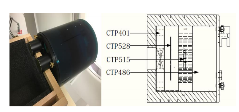
An example of physical phantoms is the Catphan® 500 (The Phantom Laboratory, Salem,
NY). This phantom is widely used for testing the performance of CT scanners. The phantom consists
of five modules to assess the image quality; (i) CTP401 for slice width, sensitometry and pixel size,
(ii) CTP528 for line pair and point source spatial resolution, (iv) CTP515 for sub slice and supra
slice low contrast and (v) CTP486 for image uniformity.
The researchers state that the Lungman anthropomorphic chest phantom is equipped with a phantom that mimics the heart, with surrounding structures and tissues very similar to a real patient. The heart features are lacking, however, with simulation allowed through only one, homogeneous material.
“Consequently, the lack of CCTA image features can be addressed by replacing the current cardiac insert with a newly designed cardiac insert phantom that can provide appropriate CCTA image appearances similar to the real human heart,” state the researchers.
They also discuss the benefits of 3D printing in phantom development, as researchers have studied ways to make phantoms mimicking different parts of the anatomy. So far, however, a 3D printed insert of a Lungman phantom has not been created.
“Consequently, evidence to demonstrate the application of this 3D-printed cardiac insert phantom for CCTA dose optimisation is lacking,” state the researchers.

(a) The Lungman anthropomorphic chest phantom by Kyoto Kagaku co., Japan is placed
on the CT table. (b) The removable cardiac (arrow) and lung structures contained within the
Lungman phantom.
The goals of the study include:
- 3D printing a cardiac insert phantom created from volumetric CT image datasets
- Investigating the 3D printed phantom in evaluation an IR algorithm
- Evaluating optimal IR algorithm strengths for low-tube voltage CCTA protocols
We have seen 3D printing in a wide range of medical models, from those fabricated to train medical students to those meant to streamline patient care, and models created for pre-surgical planning. Not a lot of research has been involved in creating cardiac phantoms, though. For this study, they were able to 3D print an insert with the same specifications as the first Lungman cardiac insert:
“The new 3D printed cardiac insert was positioned into the Lungman phantom and scanned using a standard CCTA protocols. The resultant images were compared to the patient and Catphan® 500 phantom images. HU values of the attenuating materials within the new 3D-printed cardiac insert phantom were comparable to tissues in the patient image datasets and materials in the Catphan® 500 phantom.”
The insert was created on a Creatbot DM Plus 3D printer. Filling materials like contrast media, oil, water, and jelly were loaded into the phantom insert.
“The 3D-printed cardiac insert phantom was positioned within the anthropomorphic chest phantom and thirty repeated CT acquisitions performed using a multi-detector scanner at 120-kVp tube potential. Attenuation (Hounsfield Unit, HU) values were measured and compared to the image datasets of realpatient and Catphan® 500 phantom,” stated the researchers in their paper.
The research team found that using an IR algorithm does permit lower exposure through reducing image noise during image reconstruction.
“The results of our analysis showed that all types of IR algorithms significantly reduce radiation dose with no significant difference in diagnostic image quality between FBP and IR algorithm,” stated the team.
Ultimately, they discovered that 3D printing was suitable for dose optimization studies, allowing for investigation of IR algorithm on dose reduction.
“Evidence provided should also provide new horizons to researchers for novel 3D-printed phantoms and facilitate better CT optimisation process regarding their clinical implementation,” concluded the researchers.
What do you think of this news? Let us know your thoughts! Join the discussion of this and other 3D printing topics at 3DPrintBoard.com.

(a) The Catphan® 500 phantom (The Phantom Laboratory, Greenwich, NY, USA); (b) The
3D-printed cardiac insert phantom; (c) The Catphan® 500 phantom was positioned on the scannertable; and (d) The anthropomorphic chest phantom (Lungman N-01, Kyoto Kagaku, Japan), with the 3D-printed cardiac insert phantom positioned within (arrow), was placed on the scanner table.
Subscribe to Our Email Newsletter
Stay up-to-date on all the latest news from the 3D printing industry and receive information and offers from third party vendors.
You May Also Like
Further Understanding of 3D Printing Design at ADDITIV Design World
ADDITIV is back once again! This time, the virtual platform for additive manufacturing will be holding the first-ever edition of ADDITIV Design World on May 23rd from 9:00 AM –...
3D Printer Maker EVO-tech Reborn as NEVO3D — Once More With Feeling
EVO-tech was a 3D printing service and original equipment manufacturer established in 2013 and based in Schörfling am Attersee, Austria. The company produced high-quality material extrusion systems featuring linear bearings,...
3D Systems Brings 3D Printed PEEK Cranial Implant to the U.S. with FDA Clearance
For more than 10 years, 3D Systems (NYSE:DDD) has worked hand-in-hand with surgeons to plan over 150,000 patient-specific cases, and develop more than two million instruments and implants from its...
CDFAM Returns to Berlin for Second Annual Symposium
The second CDFAM Computational Design Symposium is scheduled for May 7-8, 2024, in Berlin, and will convene leading experts in computational design across all scales. Building upon the first event...