3D Printing the Left Atrial Appendage for Better Closure Procedures in Surgery


Mia von Knorring, Areli Reyes, and Brandon Mukai recently submitted ‘Left Atrial Appendage Printing Procedure’ to the Faculty of the Biomedical Engineering Department at California Polytechnic State University – San Luis Obispo as a senior project. Exploring the use of 3D printed medical models for better visual aids in cardiac procedures, the research students aimed to help with the closing off the left atrial appendage.
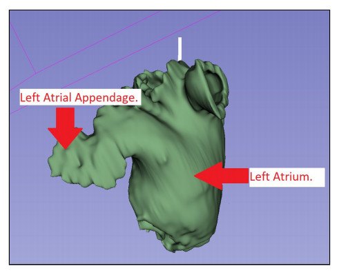
Virtual model of the left atrium and left atrial appendage
The research is centered around assisting in the Watchman left atrial appendage closure procedure—a surgery only 100 surgeons in the US are trained for, meant to eliminate debris from the left atrial appendage that could cause a stroke. With a detailed 3D model, cardiac physiologists are better able to size and position the device. The study began with alterations to the original algorithm by Dr. Chris Porterfield, and then a final 3D design taken from CT scans, segmented, and 3D printed with PLA and PVA on an Ultimaker 3D printer (with a final cost of $118.94 after tax and shipping).
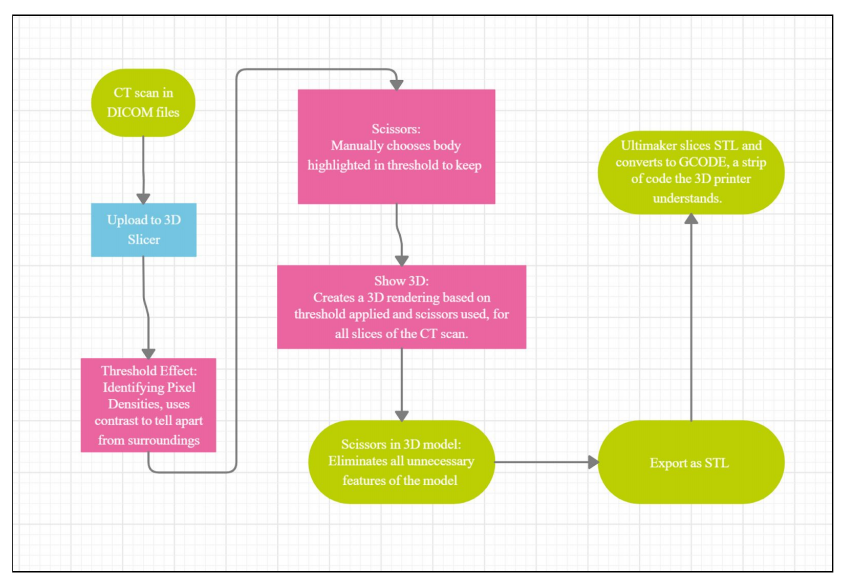
Manufacturing Process flowchart

Labeled views
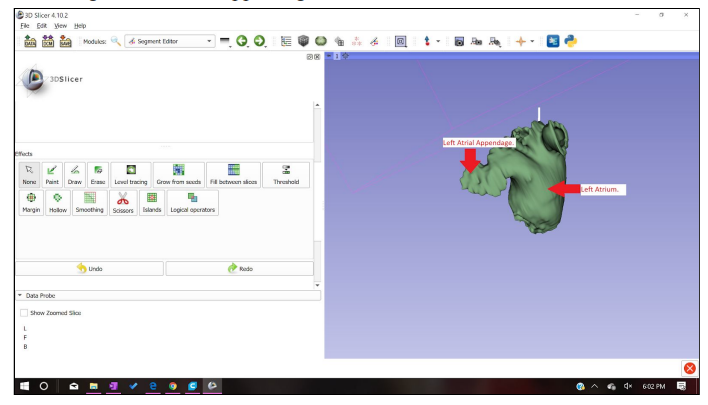
Cropped 3D model
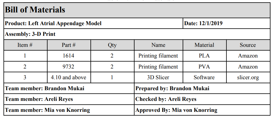
Bill of Materials
This is not just a research study exploring feasibility, but it explains how to create the 3D model from beginning to end, using 3D slicer—explaining the proper use of pixel density and understanding variances based on CT scan parameters. Ultimately, the research students explained that they would have included a more expanded threshold range, allowing for customizations depending on the left atrium and appendage.
They also added that in later use, 3D printing materials that are more like tissue would be an improvement:
“More flexible material is clinically relevant and could allow doctors to see the compression the Watchman could cause on the appendage to ensure that the device is correctly sized. Many sections of the procedure create variability that could change procedure outcomes,” stated the authors.
“We had the same lab assistant print all of our models, but he created certain parameters and printer features for all his prints that could possibly change between operators. To mitigate this risk, we recorded all notable parameters in our procedure. Variability could also arise when anatomy is being cropped. Operators could remove too much necessary anatomy or include more anatomy to create a larger model. In some patients, the pulmonary arteries are near the left atrial appendage.”
The authors make it clear too that these types of 3D slicing and printing are valid for a wide range of other applications and in other cardiac exercises such as sizing heart valves.
Guidelines for use of the model are included in the paper, and a request for user feedback, which so far has been taken by both Dr. Porterfield and his clinical specialist Sarah Griess.
“All comparisons had a p-value<.05, confirming repeatability and reproducibility of these features> confirming repeatability and reproducibility of these features. Depth measurements however resulted in a p-valve of .036 between operators. This discrepancy could be due to the challenging angle measurement,” concluded the researchers.
“Since the anatomical models are curved and irregularly shaped, it was difficult to measure the depth consistently between models. Lack of consistency PVA removal could also cause this design specification failure. On some models, PVA still remained in small cavities after post processing was completed even though we tried to remove as much as possible. Even though inconsistency between operators exists in the depth measurement, depth is not a critical measurement to Watchman sizing.”
Overall, the models were verified as accurate.
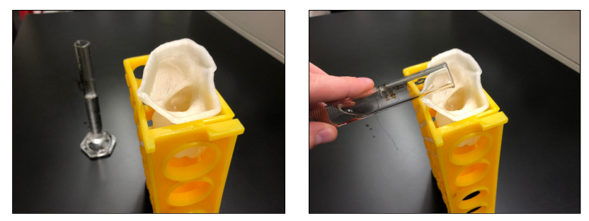
Volume measurement set-up
Today, 3D printing models abound within the medical arena for use in diagnosis, treatment, surgical planning, and more. What do you think of this news? Let us know your thoughts! Join the discussion of this and other 3D printing topics at 3DPrintBoard.com.
[Source / Images: ‘Left Atrial Appendage Printing Procedure’]
Subscribe to Our Email Newsletter
Stay up-to-date on all the latest news from the 3D printing industry and receive information and offers from third party vendors.
Print Services
Upload your 3D Models and get them printed quickly and efficiently.


You May Also Like
DREAMing in Dayton: DREAM Symposium Covers AM, AI, Supply Chain, & More
This month, I attended a manufacturing industry event, like I often do. But instead of getting on a plane to New York City, or driving four hours to Youngstown, I...
ORNL Origami Creates Large Foldable Structures
Oak Ridge National Laboratory (ORNL) is using a hybrid 3D printing method to make foldable panels. At the Department of Energy’s (DOE) Manufacturing Demonstration Facility (MDF) at ORNL, researchers turned...
Phase3D’s In-Situ Monitoring Lands $2.9M in Oversubscribed Round
The use of metal additive manufacturing (AM) for production at scale appears to be steadily increasing, as evidenced by recent announcements like EOS’s sale of 30 M4 ONYX systems to...
Excellent Desktop Injection Molding, Made in Italy by Robot Factory
I was captivated when I saw my first Robot Factory 3D printer. The robust, precise machine was built to last. And this was in an era of very flimsy, disposable,...