
China: 3D Printed Models Aid in Treatment & Surgery for Hip Fractures


Researchers from the Department of Orthopedics Surgery, The Second Affiliated Hospital, and Yuying Children’s Hospital of Wenzhou Medical University have recently published a study regarding a more effective approach for hip fractures. In ‘3D printing-based Ganz approach for treatment of femoral head fractures: a prospective analysis,’ the authors explore preferred treatment of femoral head fractures (not a common injury), and the inherent challenges and controversies—along with offering recommendations for improved methods via 3D printing technology.
![]()
For the patient stricken with a femoral head fracture—one occurring at the highest part of the thigh bone—surgery is complex and can be difficult, leading to many different issues later too. In this study, 17 patients were assessed over a five-year period from 2012-2017, with 3D printed models created for treatment and surgical simulation.
Each of the patients had been admitted to the emergency room initially and were assessed by the Adult Trauma Life Support (ATLS™) guidelines.
“There was a patient with irreducible dislocation with the femoral head buttonholed through the capsule and impaled against the posterior rim of the acetabulum. This patient indicated open reduction and internal fixation with emergency surgery; thus, the patient was excluded due to the lack of time for 3D printing preoperative,” explained the researchers regarding the subjects of their study.
“All the rest [of the] patients received a CT scan to evaluate the quality of reduction. Operative treatment was indicated for fractures displaced 2 mm or more in CT scan. Two patients were lost to follow-up, and two patients were excluded because of follow-up of less than 2 years, leaving a total of 12 patients as the study population.”
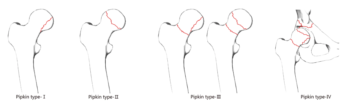
Pipkin classification of femoral head fractures with posterior hip dislocations. Types I and II are distinguished by the position of the fracture in relation to the fovea. Type I is below the fovea with the fracture outside of the weight-bearing joint-parts, whereas type II fractures involve the more cranial, weight-bearing parts. Type III is any fracture of the head in combination with a femoral neck fracture. Additional fractures of the acetabulum are classified as type IV.
CT data was used to create 3D models of each patient’s fracture, making the details of each one clear. The researchers were able to complete trochanteric osteotomies, along with simulating intraoperative reduction and fixation techniques using the models. The authors stated that they were also able to choose all the proper hardware for the surgery, using the life-size 3D printed model of the hip, along with outlining length, location, and orientation. The model could also be used as a surgical guide from the operating room.
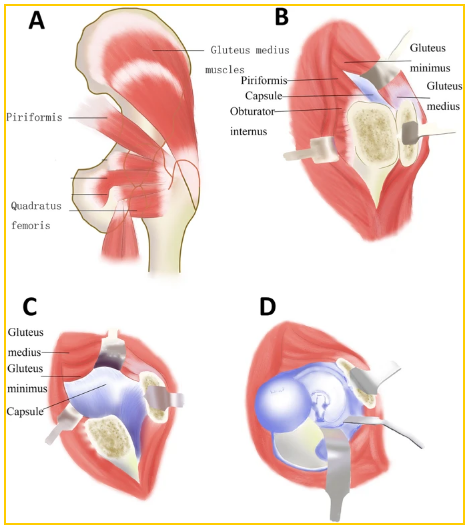
Osteosynthesis of a femoral head fracture using the Ganz approach. a The position of the trochanteric osteotomy: The osteotomy exits just anterior to the most posterior insertion of gluteus medius proximally and the entire origin of vastus lateralis remains on the trochanteric fragment distally. b After the osteotomy of the trochanter, the osteotomized trochanter fragment, including the tendon of gluteus minimus, is reflected anteriorly. Through blunt separation, the tissue space between the tendon of piriformis and gluteus minimus is enlarged and gluteus minimus contracts upward, which contributes to adequately expose the capsule. c Make a Z-shaped capsule incision to further rotate outward and flex the femur. d After dislocation of the femoral head, lower the knee and rotate the affected leg, which can make the fracture can easily visualized, reduced, and stabilized.
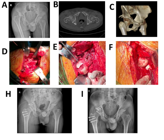
This was a 58 year old male with type IV fracture caused by car accident. a The femoral head fracture, posterior dislocation of the hip, and posterior acetabular wall fracture. b, c The CT scan after reduction which showed detail of the fracture. d The osteotomy of the trochanter: the osteotomized trochanter fragment is reflected anteriorly. e, f The reduction and fixation with femoral head fracture and acetabular fracture. h The X-ray after operation. i The X-ray of 1 year after operation, which showed the fracture has healed and there is no AVN.
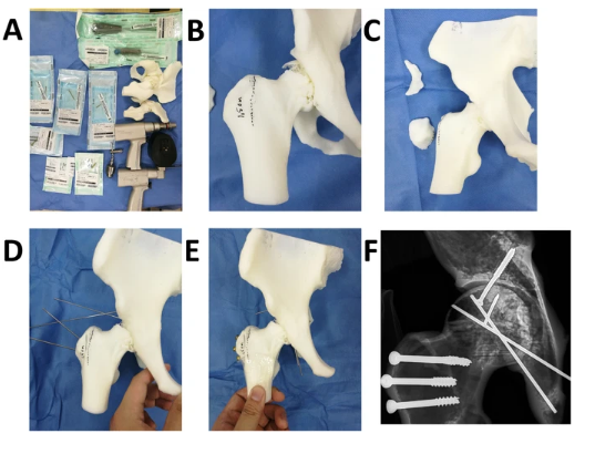
Simulation of the surgery in 3D printing model for the patient of Fig. 3. a Preparation of simulative surgery. b Mark at the osteotomy line. c Complete the femoral trochanter osteotomy and separate the fracture fragment of the posterior wall of the acetabulum. d, e Reduction and fixation with suitable length screws and Kirschner wire. f X-ray after the simulative surgery.
Mean operative time for all procedures in the study was 124.2 ± 22.1 min, and estimated blood loss was 437.5 ± 113.1 ml, in comparison to other studies demonstrating surgical time of 21 min for Type I or II fractures and 195 min for Type IV fractures, and blood loss of 1334 ml in the isolated femoral head fractures and 1557 ml in head and acetabulum fractures. The authors confirmed that with the use of the 3D printed model, they were able to reduce time in the operating room, and blood loss, but they do agree that further study is required.
Six patients had an excellent result, four had good results, and two had poor results—all of which the authors state is comparable to other recent studies with ‘good outcomes.’
“Our study revealed that 3D printing-based Ganz approach could obtain excellent surgical exposure and protection of the femoral head blood supply, reduce the operation time and intraoperative blood loss, make the precise osteotomy, and anatomically reduce and fix the intra-articular fragments, which provided satisfactory results of treatment in femoral head fractures,” concluded the researchers.
“At the same time, the probability of AVN in this surgical method was much lower than others and the impact on MCFA was avoided with the 3D printing-based Ganz approach.”
The use of 3D printed models in the treatment of patients today is not only incredibly fascinating in the amount of detail that can be uncovered, but also in the benefits offered to both patients and surgeons for other types of procedures too like renal surgeries, chest wall reconstruction, eye socket repair, and so much more.
What do you think of this news? Let us know your thoughts! Join the discussion of this and other 3D printing topics at 3DPrintBoard.com.

Subscribe to Our Email Newsletter
Stay up-to-date on all the latest news from the 3D printing industry and receive information and offers from third party vendors.
Print Services
Upload your 3D Models and get them printed quickly and efficiently.


You May Also Like
The Next Phase of EB-PBF Will Be Defined by Beam Control
The bar for metal additive manufacturing has moved. Early on, the question was often simple: Can the machine print the material and produce a dense part? That still matters, but...
UAS Additive Strategies Shows How Fast Drone Manufacturing Is Changing
The recent UAS Additive Strategies online event, hosted by 3DPrint.com and Additive Manufacturing Research (AM Research), brought together leaders from across the additive manufacturing (AM) and drone industries to discuss...
The Drone Industry is Showing Where 3D Printing Delivers Real Value, AM Research Report Finds
The rapid rise of drones is creating one of the biggest opportunities for additive manufacturing (AM). Whether they’re used on battlefields, inspecting bridges or crops, or delivering supplies, drones need...
The Longevity Economy Needs a Factory
Longevity has become one of the biggest stories in healthcare. Every week seems to add a new announcement about an anti-aging therapy, an AI-powered drug discovery platform, or a startup...


