Zhejiang University Sheds Light on APVC with 3D Printed Surgical Models


Researchers from China’s Zhejiang University are looking for new ways to improve preoperative planning for procedures for children, with their findings outlined in the recently published ‘Utility of three-dimensional printing in preoperative planning for children with anomalous pulmonary venous connection: a singer center experience.’ Many of the greatest benefits were put into action for this study as the 3D printed models could be used for completing diagnosis, assessing treatment options, and planning for surgery.
![]() In this study, Chinese medical scientists studied 17 children diagnosed with anomalous pulmonary venous connection (APVC) from November 2017 to January 2019. Ages ranged from only two days old to twenty months old, with the following variations:
In this study, Chinese medical scientists studied 17 children diagnosed with anomalous pulmonary venous connection (APVC) from November 2017 to January 2019. Ages ranged from only two days old to twenty months old, with the following variations:
- Ten children suffering from total supracardiac APVC
- One child suffering from intracardiac APVC
- Mixed type APVC in one child
- Partial APVC in three children
Data from CT scans was imported into Mimics 19.0 software for 3D modeling and design of the heart model to display elements of the heart such as papillary muscles, muscle bundles, and outflow tracts. While very little research has been performed for APVC citing the assistance of 3D printing, it is clear from this study that the use of models allows for much greater light to be shed on the condition.
“We manually labelled each area according to the left ventricle (LV), right ventricle (RV), LA, RA, aorta (AO), and pulmonary artery (PA) area modules of the CT heart module and distinguished them with different colors. Special attention was paid to labeling the boundaries of each part,” stated the researchers.
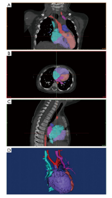
Image segmentation and postprocessing in Mimics 19.0 software. The colored masks were segmented for 3D modeling. (A) Coronal plane; (B) transverse plane; (C) Sagittal plane; (D) 3D modeling. Green, superior vena cava and right atrium; purplish red, pulmonary veins and left atrium; purple, right ventricle; orange, left ventricle; red, aorta; dark blue, pulmonary artery.

Four cases diagnosed with supracardiac type TAPVC. (A) Refers to patient 1, view from posterior; (B) refers to patient 5, view from posterior; (C) refers to patient 9, view from posterior; (D) refers to patient 10, view from posterior. *, obstruction exists at the junction of the common pulmonary venous confluence to the left-sided vertical vein (VV). Ao, aorta; PA, pulmonary artery; PV, pulmonary vein; SVC, superior vena cava. Orientation labels: I, inferior; L, left; R, right; S, superior.

Two cases diagnosed with intracardiac type TAPVC. Part of the hollowed heart model was segmented to emphasize the PV and RA we focused on. Pulmonary vein through the coronary sinus opening in the right atrium. (A) Refers to patient 11, view from inferior; (B) refers to patient 12, view from the right. CS, coronary sinus; IVC, inferior vena cava; PV, pulmonary veins; RA, right atrium. Orientation labels: A, anterior; I, inferior; L, left; P, posterior; R, right; S, superior.
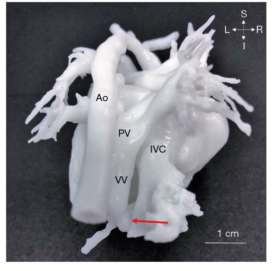
One case (patient 13) diagnosed with infracardiac type TAPVC, view from posterior. The red arrow indicates the junction of the pulmonary vein and the inferior vena cava. Ao, aorta; IVC, inferior vena cava; PV, pulmonary vein; VV, vertical vein; TAPVC, total anomalous pulmonary venous connection. Orientation labels: I, inferior; L, left; R, right; S, superior.
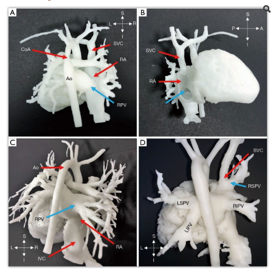
Three cases diagnosed with PAPVC. Images (A,B) both represent patient 15. (A) The blue arrow points to RPV flowing into the right atrium. View from posterior. (B) The blue arrow represents the outlet of RPV, flowing into the right atrium. View from the right. (C) Patient 16 diagnosed with PAPVC; the blue arrow represents RPV flowing into the right atrium. View from posterior. (D) Patient 17. The blue arrow represents RSPV flowing into the SVC. View from posterior. Ao, aorta; CoA, coarctation of aorta; IVC, inferior vena cava; LIPV, left inferior pulmonary vein; LSPV, left superior pulmonary vein; RA, right atrium; RIPV, right inferior pulmonary vein; RPV, right pulmonary vein; RSPV, right superior pulmonary vein; SVC, superior vena cava. Orientation labels: A, anterior; I, inferior; L, left; P, posterior; R, right; S, superior.
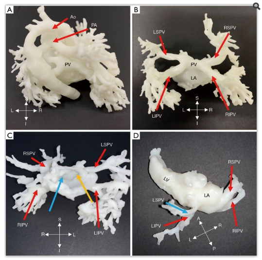
Two cases underwent cardiac CT examination during follow-up. (A,D) Blood volume; (B,C) hollowed models. Images (A,B,C) represent patient 3 after repaired supracardiac TAPVC. (A,B) view from posterior; (C) view from anterior. (C) The blue and yellow arrows represent the opening of the right pulmonary vein and left pulmonary vein. There is no anastomotic stenosis. (D) Superior view of patient 4 after repaired supracardiac TAPVC. Stenosis of LSPV (blue arrow) is shown compared to other pulmonary vein branches. Ao, aorta; LA, left atrium; LIPV, left inferior pulmonary vein; LSPV, left superior pulmonary vein; PA, pulmonary artery; PV, pulmonary vein; RIPV, right inferior pulmonary vein; RSPV, right superior pulmonary vein. Orientation labels: A, anterior; I, inferior; L, left; P, posterior; R, right; S, superior.
3D printing of the personalized heart models was completed via an ISLA 650 3D printer (Shining 3D, China). Preoperative planning was then based on the models, along with medical history of the patients, and imaging data. The models were also used as surgical guides in the operating room upon being sterilized. Each patient-specific heart model took around half an hour to two hours to model, with 3D printing requiring anywhere from two to five hours. Surgeries were performed on all 17 patients, and each procedure was successful.
“The malformations demonstrated by the 3D models were consistent with intraoperative observations, and presurgical planning was in line with real surgery programs. These heart models could be sterilized and brought into the operating room for surgery navigation. These 3D models greatly assisted the presurgical planning for APVC surgery and were of great clinical value from our experience.
“After surgeries, these heart models were evaluated on whether they were of high quality, and whether they could help presurgical planning, reduce unforeseen circumstances, and benefit medical education. An evaluation pertaining to the issues above was conducted via questionnaire by our cardiac surgeons and cardiologists.”
3D printed heart models and medical devices such as implants have been used in connection with cardiac issues and defects, with guides and models used in everything from complex medical training to pediatric surgeries, methods for creating heart patches, and more.
What do you think of this news? Let us know your thoughts! Join the discussion of this and other 3D printing topics at 3DPrintBoard.com.
[Source / Images: ‘Utility of three-dimensional printing in preoperative planning for children with anomalous pulmonary venous connection: a singer center experience’]Subscribe to Our Email Newsletter
Stay up-to-date on all the latest news from the 3D printing industry and receive information and offers from third party vendors.
Print Services
Upload your 3D Models and get them printed quickly and efficiently.


You May Also Like
The New Dental Lab: “Three Technicians Can Handle a Hundred Arches,” Says Digital Dentistry Expert Josh Jakson
Josh Jakson’s path into digital dentistry started long before he had a job title. He grew up around it. His father, a Polish immigrant, started the family’s dental laboratory in...
AM Asia Watch: China’s HeyGears Lands $44M to Expand Beyond Dental 3D Printing
Chinese 3D printing company HeyGears raised more than 300 million Yuan (roughly $44 million) in a new Series C funding round as it looks to expand beyond its industrial and...
Asia AM Watch: China’s SHINING 3D Restarts IPO Review Process
SHINING 3D is moving forward again with its plans to go public in China, after restarting its Beijing Stock Exchange (BSE) initial public offering (IPO) review process and filing updated...
3D Printing Financials: Align’s Growth Runs on Volume
Align Technology (Nasdaq: ALGN) kicked off 2026 with steady financial results, with most of the growth coming from its core high-volume 3D printing business. The maker of Invisalign reported first-quarter...