

Canada: University Researchers 3D Print GlioMesh to Treat Brain Cancer


In the recently published ‘A Drug-Eluting 3D-Printed Mesh (GlioMesh) for Management of Glioblastoma,’ Canadian researchers take on the topic of using 3D printing for better treatment of glioblastoma (GBM) as current surgical procedures, radiation therapy, and medications still do not seem to be making an impact on survival rates.
In this study, the researchers offer a new method for treatment, via GlioMesh, made from 3D printed hydrogels that are filled with temozolomide microparticles (TMZ). GBMs are one of the most aggressive forms of brain cancer, affecting nearly 50 percent of patients with brain tumors within the US. Today, there is less than a ten percent chance of survival over five years for patients diagnosed with GBM.
“The standard therapy for GBM is maximal safe surgical resection, followed by radiation and chemotherapy with temozolomide (TMZ) for 6 months,” explain the researchers. “After the radiotherapy is finished, the monthly administration of TMZ is maintained for 6 months up to one year. Together, these typically add only months of additional survival. Even with the current advances in microsurgical techniques, tumor recurrence is the norm, typically occurring within 1–2 cm of the original tumor border.”
Obstacles in treating/managing GBM include:
- Issues with complete removal of the tumor, often spread in ‘finger-like projections’
- Ineffectiveness of chemotherapy to treat areas deep within brain tissue
- Challenges due to the blood-brain/tumor barrier
- Drug-resistant characteristics of GBM cancer stem cells
The researchers are striving to:
- Increase the amount of survival time
- Increase long-term survival rate
- Improve quality of life for patients
While TMZ can overcome the blood-brain barrier, high doses are often required, and side effects can be brutal; however, the researchers explain that is it possible to bypass some of these challenges with localized delivery of the drug. With the drug-releasing mesh developed for this study (consisting of alginate hydrogel and laden with TMZ‐loaded PLGA microspheres), the researchers found they could release TMZ over the tumor site for seven weeks at a time.
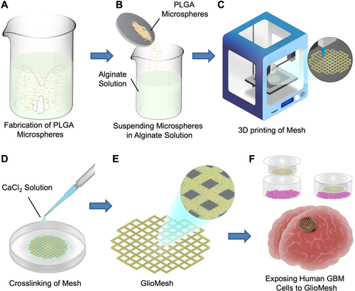
Schematic demonstration of GlioMesh and its fabrication process. A) O/O emulsion solvent evaporation technique for fabrication of TMZ‐loaded PLGA microspheres with high encapsulation efficiency. B) Preparation of bioink. C) 3D printing of alginate mesh containing TMZ‐loaded PLGA microspheres. D) Cross‐linking of the printed mesh. E) 3D printed mesh, laden with TMZ‐releasing PLGA microspheres. F) The efficacy of GlioMesh in treatment of GBM was evaluated by various studies on U251 and U87 human GBM cells.
“Fabrication of a porous mesh by 3D printing is an enabling technology that offers the advantages of higher mass transport of the drug to the surrounding tissue due to higher surface to volume ratios, better cellular infiltration, and enhanced delivery of nutrients and oxygen to the underlying tissue,” explained the researchers.
The study showed poor ‘encapsulation efficiencies’ of 0.87 ± 0.52%, and 1.34 ± 0.03% for the microspheres prepared with O/W and W/O/W emulsion, respectively. This had also been the result for previous researchers engaging in similar work. Further refinements did not show much of an improvement.
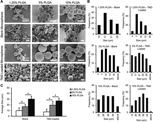
Characterization of PLGA microspheres prepared with different PLGA concentrations. A) SEM images of blank and TMZ‐loaded PLGA microspheres prepared with 1.25%, 5%, and 10% PLGA concentration. Scale bars are 500 µm. B) Size distribution of blank and TMZ‐loaded PLGA microspheres fabricated with various PLGA concentrations. C) Average size of blank and TMZ‐loaded PLGA microspheres prepared with 1.25%, 5%, and 10% PLGA concentration. Increase in PLGA concentration resulted in fabrication of microspheres with larger average diameter. Each data point represents the average ± SD. *p < 0.0005.
As the researchers increased print-head pressure, they were finally able to deposit more alginate, increasing fiber diameter. Greater control over the 3D printed meshes occurred with the proper amount of viscosity. Adding polymeric microspheres also helped encourage longer TMZ release at the tumor.
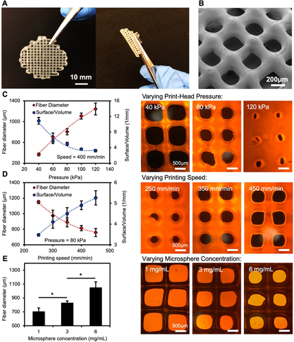
Characterization of 3D bioprinted alginate mesh. A) Photographic images of TMZ‐releasing alginate mesh. Right image shows the flexibility of GlioMesh, which is a suitable feature for a brain implant to comply with the underneath irregularly shaped tissue. B) SEM image of GlioMesh showing its porous structure. C) The effect of print‐head pressure on the fiber characteristics. Higher pressure on the nozzle resulted in larger fiber diameter and smaller surface‐to‐volume ratio. Microscopic images of alginate mesh printed with 40, 80, and 120 kPa pressure. D) The effect of printing speed on the characteristics of the 3D bioprinted fiber. Printing with higher speed resulted in decreased fiber diameter and increased surface‐to‐volume ratio. Microscopic images of alginate meshes printed with 250, 350, and 450 mm min−1 of printing speed. E) The effect of microsphere concentration on the fiber diameter in the 3D printed meshes. Fiber diameter increased by higher microsphere densities. Microscopic images of GlioMesh printed with 1, 3, and 6 mg mL−1 of microsphere concentration. Each data point represents the average ± SD with n = 6. *p < 0.005 and **p < 0.0005.
“GlioMesh demonstrated a sustained release of TMZ over 56 days, which circumvents the need for frequent oral administration of this chemotherapy drug in GBM patients,” concluded the researchers. “GlioMesh showed superior cytotoxic effect over free TMZ due to the preservation of the drug from degradation over the course of treatment and maintaining the level of autophagy in GBM cells.
“Furthermore, higher degree of mitochondrial damage was achieved by sustained delivery of TMZ in comparison with free TMZ. All in all, GlioMesh holds great promise in the management of GBM by reducing the side effects of chemotherapy, circumventing the BBB and associated challenges, and providing more flexibility toward using a combinational therapy approach that is tailored to each patient.”
The amount of cancer research and treatment today that includes 3D printing in its successes is staggering—from microfluidic devices to the printing of tumors, microtumors, and more for assistance in critical research.
What do you think of this news? Let us know your thoughts! Join the discussion of this and other 3D printing topics at 3DPrintBoard.com.
[Source / Images: ‘A Drug-Eluting 3D-Printed Mesh (GlioMesh) for Management of Glioblastoma’]Subscribe to Our Email Newsletter
Stay up-to-date on all the latest news from the 3D printing industry and receive information and offers from third party vendors.
Print Services
Upload your 3D Models and get them printed quickly and efficiently.


You May Also Like
3D Printed Chip Packaging Specialist XTPL Enters Japanese Market
The additive manufacturing (AM) industry naturally wants to move beyond prototyping to production at scale, and the industry is certainly starting to demonstrate success with that objective, especially in Asia....
UT Researchers Use 3D Printing to Develop “Tabletop EUV Lithography” Process
Photolithography, the semiconductor manufacturing process whereby lasers transfer patterns onto chemical layers coating a substrate, is one of the most amazing industrial processes humanity has ever created. It is also...
3D Printing News Briefs, May 30, 2026: RIMPAC 2026, Acquisition, Ceramic Implants, & More
We’re kicking things off with materials news in this weekend’s 3D Printing News Briefs. Then it’s on to a hybrid manufacturing system for a maritime exercise, an expansion of industrial...
The University of Utrecht: “3D Printing Could Change Who Gets to Become a Manufacturing Power”
For decades, manufacturing has mostly been controlled by countries with huge factories, lower labor costs, and industrial systems that took years, sometimes decades, to build. But Utrecht University human geographers...


