

3D Printed Medical Models Give Better Preoperative Education to Aneurysm Patients


In ‘Obtaining Informed Consent Using Patient Specific 3D Printing Cerebral Aneurysm Model,’ Korean researchers delve into an area that is becoming more well-known as a benefit of 3D printing, but not one that is discussed as much as more complex medical procedures and technological advances are—in regards to the helpful education that can be afforded to the patient before surgery.
While doctors and students are provided with invaluable tools via 3D printing to pave the way for patient-specific treatment, the same models they are using can also be advantageous to their patients and their families, all of whom may be struggling to come to grips with a life-threatening health issue and impending surgery—along with trying to make the decision whether to go forward with a procedure or not.
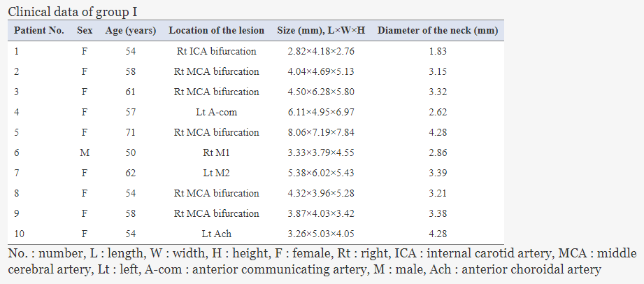
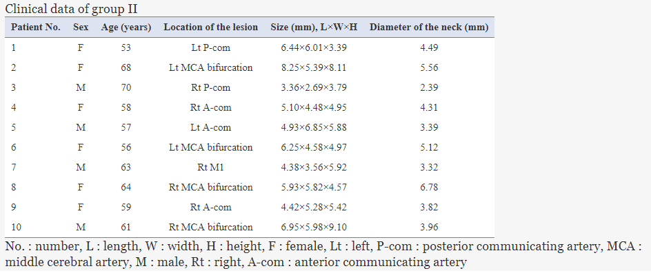
For this study (approved by the Pusan National University Hospital Institutional Review Board), 20 patients with unruptured intracranial aneurysms were divided into two groups. Ten patients were educated via the 3D printed model (created on the Nobel 1.0A with PLA), while ten others were given the traditional computed tomography angiographies (CTA).
The patients in the study were also asked to fill out a questionnaire regarding not only their satisfaction with the surgery—but also their comprehension level thanks to the model and explanation. These models allow patients and doctors, as well as junior surgeons and other students, to understand more about the positional relationship between the aneurysms and arteries, along with:
- Branches
- Bones
- Brain parenchyma
- Cranial nerves
All 20 patients received identical information about aneurysm physiology, the medical procedure, and the risk involved.
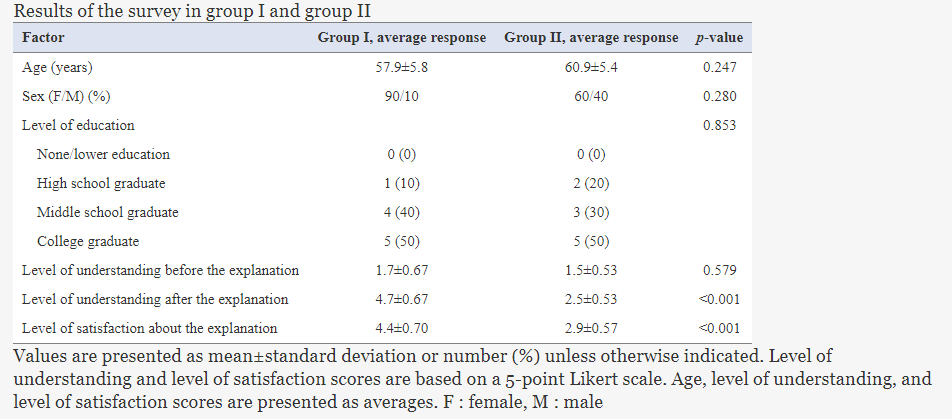
A single doctor was not only responsible for all 20 surgeries, but also for the explanation of the procedure at hand—removing any concerns about bias regarding the educational process, or differences in the procedure. The model was used as an accompaniment to the preoperative consent, with the study proving to be conducive to a clear understanding of the patients’ understanding of the process due to new technology. Upon reviewing the results of the research, the authors found the data to suggest that the 3D printed models significantly improved the process, as patients had better understanding of what was happening, along with higher satisfaction regarding the surgery.
“The main limitation of 3D printing technology is that building a 3D model involves a 3D rendering and 3D printing, which are time consuming and costly. However, with the development of newer printers and the broadening of printed materials, the time and cost have been reduced. In this study, we used the Nobel 1.0A (XYZPrinting), which is small (280×345×590 mm) and lightweight (9.6 kg),” concluded the researchers. “The process time for making the 3D models is 3.7 hours on average. The material cost of each model is approximately 3500 Korea won. The time and cost will decrease as 3D printing technology advances. As 3D printing technology progresses and costs fall, patient-specific 3D printing may become standard for both clinical and educational purposes.
“Given that this was only a single-center experience and that there were only 10 patient-specific models used to evaluate patient understanding, additional larger prospective studies will be needed to confirm the usefulness of a patient-specific 3D print model for patient understanding and satisfaction with aneurysm clipping surgery.”
3D printed models have not only changed the face of medicine when it comes to diagnosing and understanding serious health conditions like cerebral aneurysms or brain tumors, but also in terms of training, allowing doctors and medical students to understand more about unusual or completely new procedures, and rely on models as surgical guides.
What do you think of this news? Let us know your thoughts! Join the discussion of this and other 3D printing topics at 3DPrintBoard.com.
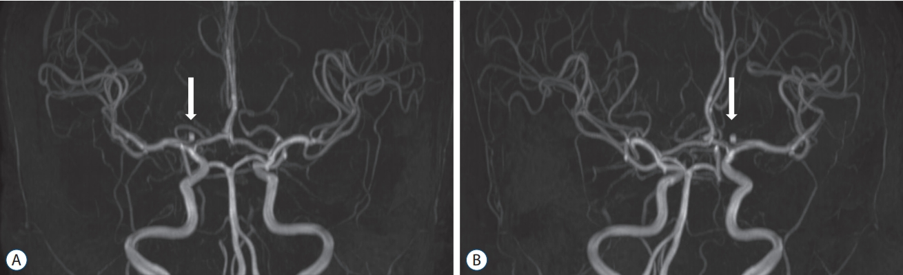
Magnetic resonance angiography (MRA) of a right internal carotid artery bifurcation aneurysm before surgery. White arrows showed aneurysms on MRA. A : Anteroposterior view. B : Posteroanterior view.

Three-dimensional reconstruction of a right internal carotid artery bifurcation aneurysm before surgery using Materialise Mimics software (Materialise, Leuven, Belgium). A : Anteroposterior view. B : Posteroanterior view.
Subscribe to Our Email Newsletter
Stay up-to-date on all the latest news from the 3D printing industry and receive information and offers from third party vendors.
Print Services
Upload your 3D Models and get them printed quickly and efficiently.


You May Also Like
3D Printed Chip Packaging Specialist XTPL Enters Japanese Market
The additive manufacturing (AM) industry naturally wants to move beyond prototyping to production at scale, and the industry is certainly starting to demonstrate success with that objective, especially in Asia....
UT Researchers Use 3D Printing to Develop “Tabletop EUV Lithography” Process
Photolithography, the semiconductor manufacturing process whereby lasers transfer patterns onto chemical layers coating a substrate, is one of the most amazing industrial processes humanity has ever created. It is also...
3D Printing News Briefs, May 30, 2026: RIMPAC 2026, Acquisition, Ceramic Implants, & More
We’re kicking things off with materials news in this weekend’s 3D Printing News Briefs. Then it’s on to a hybrid manufacturing system for a maritime exercise, an expansion of industrial...
The University of Utrecht: “3D Printing Could Change Who Gets to Become a Manufacturing Power”
For decades, manufacturing has mostly been controlled by countries with huge factories, lower labor costs, and industrial systems that took years, sometimes decades, to build. But Utrecht University human geographers...


