

Stanford University Researchers Create Better Cardiac Catheter Devices with 3D Printing


A heat map of the electrical activity of a heart projected onto a computer-simulated model of the right atrium.
The heart disorder atrial fibrillation, or AFib, is the most common rhythm disorder, and causes patients to have irregular, often rapid heartbeats, which messes up blood flow from the heart to the rest of the body. AFib affects over 6 million Americans, and causes more than 750,000 hospitalizations every year. While some AFib patients are lucky enough to have only a few problems caused by the disorder, others are not so lucky, and suffer serious complications that can require medications or even surgery.
 Surgeons use cardiac catheter devices to map a heart’s electrical activity, which can also be used to detect rhythm disturbances in a patient’s heartbeats. But these devices are often only one size, which makes it hard to catch these irregular heartbeats due to missed signals and spotty connections. A team of researchers from Stanford University is working to find new ways to build customized cardiac devices, and they’re using 3D printing to do it.
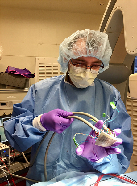
Second-year medical student Kevin Cyr, who has a background in bioengineering, said, “I’m using 3D-printed tools to design cardiac-mapping catheters, devices used by surgeons to map the electrical activity of the heart and find disturbances.”
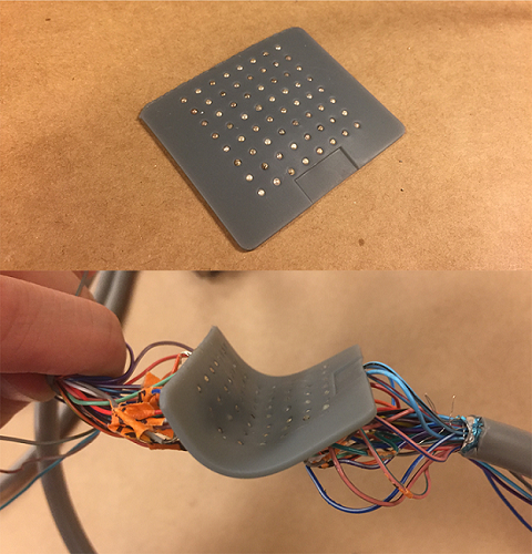
The devices are small, thin and flexible silicone membranes with tiny holes in a grid-like formation, each holding an electrode.
It’s been a few years since we’ve seen anything about 3D printed cardiac catheters, which hopefully means that 3D printing the devices has only improved with time. Stanford’s research on surveying electricity in the heart began a few years ago under Anson Lee, MD, assistant professor of cardiothoracic surgery. Cyr was interested in creating medical technologies that are able to move from research to clinical practice, and joined the team last year.
Existing one-size-fits-all cardiac catheters use electrodes to contact the heart’s surface and measure its electrical activity. The team kept the electrodes, but customized the devices to fit each individual’s heart by recording an image file of the heart during an MRI or CT scan.
“We can replicate that natural geometry and anatomy specific to that patient,” Cyr explained.
This anatomy and geometry can then be applied to a custom, 3D printed device – a thin, flexible silicone membrane with a grid of holes, each of which holds a small electrode. The 3D printed devices can very precisely track electrical activity over a specific region once it’s placed on the heart atrium’s surface.
“We can map in perfect detail this rectangular grid of information and not have to worry about missing signals, poor contact or things like that, which otherwise might throw out errors,” Cyr explained.
The data the device records is sent to a computer, which then creates a recording depicting electrical activity at certain regions. The recordings are used to make a heatmap of this activity, which surgeons then rely on to determine which regions require treatment.
While Stanford’s 3D printed cardiac catheter devices are only used on the exterior layer of the heart at the moment, the researchers are working to see if they can develop them further, in order to also map the heart’s interior surface and measure rhythmic disturbances there more accurately.

Using a soccer ball, Cyr demonstrates how the device conforms to the surface of a heart.
According to Cyr, the research team will take another couple of years to refine their 3D printed cardiac catheter devices, which have yet to be tested on humans, though they have been conformed to soccer balls. While these devices are extremely helpful to the medical field, the main goal of the team’s research is to find the problem spots in human hearts where more irregular electrical activity happens, and then get rid of them in order to bring back a normal flow of electricity.
Discuss this story and other 3D printing topics at 3DPrintBoard.com or share your thoughts in the Facebook comments below.
[Images: Kevin Cyr, Stanford University]Subscribe to Our Email Newsletter
Stay up-to-date on all the latest news from the 3D printing industry and receive information and offers from third party vendors.
Print Services
Upload your 3D Models and get them printed quickly and efficiently.


You May Also Like
Conexeu Sciences Wishes to Make a Regenerative Breast Matrix Solution (and sell shares)
Conexeu Sciences wants to commercialize a regenerative breast matrix solution. For years, Australian firm BellaSeno and Lattice Medical have been working on similar solutions. In 2018, Lattice came out with...
3D Printing News Briefs, August 23, 2025: Facial Implants, Tibial Fractures, Lamps, & More
It’s all about medical in this weekend’s 3D Printing News Briefs. BellaSeno established a clinical advisory board. MedCad is donating 3D printed facial implants to war-injured Ukrainians, and researchers in...
BellaSeno’s 3D Printed Breast Implants Keep Shape with 87% Fat Volume, Avoids Silicone Risks
At a medical conference in Austin this week, a new kind of breast implant took center stage. It is not made from silicone but from a 3D printed, fully resorbable...
BellaSeno Completes Two Clinical Trials on 3D Printed Resorbable Breast Implants
German firm BellaSeno, which is focused on 3D-printed resorbable breast implants for both augmentation and lumpectomy, has completed two clinical trials for resorbable breast implants. These trials represent the first-ever...


