Heart Transplant Surgery Performed With Help of Color Coded 3D Printed Model

 There comes a day in everyone’s life when they realize that the human heart bears no resemblance to that double lobed drawing that shows up on Valentine’s Day cards and cheap tattoos. For most of us, a vague understanding of what it looks like is all the knowledge we need. However, if you are a surgeon, and you are faced with the task of performing a heart transplant, you need to know the ins and outs in a great deal more detail.
In addition to the complexity of the organ, there is also the fact that each person’s specific heart is distinct. Add to that a heart that developed with one or more congenital deformities and you can quickly see that a surgeon is facing a daunting task. Medicine has made a huge number of strides in the last decade in terms of the possibilities for examining the specific heart upon which surgery is to be performed through micro cameras and array of data gathering equipment in order to help prevent the presentation of surprises during surgery. However, each surgery is still somewhat akin to being told that there is a problem at a particular metro station and then given a limited amount of time in which to locate and fix the problem using only a map of the ‘ideal’ metro.
For this reason, when the possibility arose for utilizing 3D printed models of individual patient’s hearts as a preparatory tool, the medical community has been quick to see the benefit. An increasing number of surgeries are being performed only after both the patient and the medical team have had the opportunity to study an exact replica of the patient’s heart. This has proven to reduce time spent in surgery, to ease patient anxiety, and to hasten patient recovery.
A team of surgeons in Dublin, Ireland recently utilized a 3D printed, color coded heart model to aid them in the preparation and performance of heart transplant surgery on a 41-year-old patient suffering from multiple heart anomalies leading to end stage heart failure. In this particular case, the list of problems encountered in the heart is enough to strike fear into even those of us who don’t necessarily understand the terminology:
- right atrial isomerism
- dextrocardia
- left inferior and superior vena cava to a single atrium
- single ventricle with right ventricular morphology
- double outlet right ventricle with transposition of the great vessels
- total anomalous pulmonary venous drainage to the superior vena cava
In a clinical tone, the team published a notice in the Proceedings of The Physiological Society in which they detailed the process for creating the full color model:
“The data were obtained using a Somaton Definition AS+ 128 slice scanner. A cardiac gated contrast enhanced Commuted Tomography study was performed at 120kV with automatic mAs modulation. Images were acquired in 0.6mm slices and reconstructed using a Siemens Syngo system. Using open source 3D Slicer software, the structures of interest were segmented. The segmented images represented the bounding lumen of the heart and Great vessels. The generated 3D model was then exported as a stereolithographic (STL) file and further model optimisation was performed using Meshlab.”
The team, headed by Michelle L. Smith of the Division of Anatomy, University College Dublin, also included L. Nolke (Cardiothoracic Surgery, Mater Misericordiae University Hospital), M.K. O’Reilly (Department of Radiology, Mater Misericordiae University Hospital), Professor James FX Jones (Head of Anatomy, University College Dublin), and Dr. John Murray (Constultant Radiologist, Mater Misericordiae University Hospital).
As the final stage in the design process, the color coding was done in Z Edit Pro, bringing the total design time to just over eight hours. A further six hours were required for printing the model on a Z Print 250 binder jetting printer. After further verification of the accuracy of the model, it was then used by the surgical team to prepare for surgery and initial reports indicate that the medical team found the model to be extremely helpful.
We’re still a ways away from printing new hearts at the press of a button, but with the introduction of 3D printing technology as a regular part of surgical practice, we’re still seeing an enormous medical benefit resulting from the technology.
Do you know of other cases where a 3D printed medical model has helped a procedure? Let us know your thoughts in the Color Coded Heart Model forum thread over at 3DPB.com.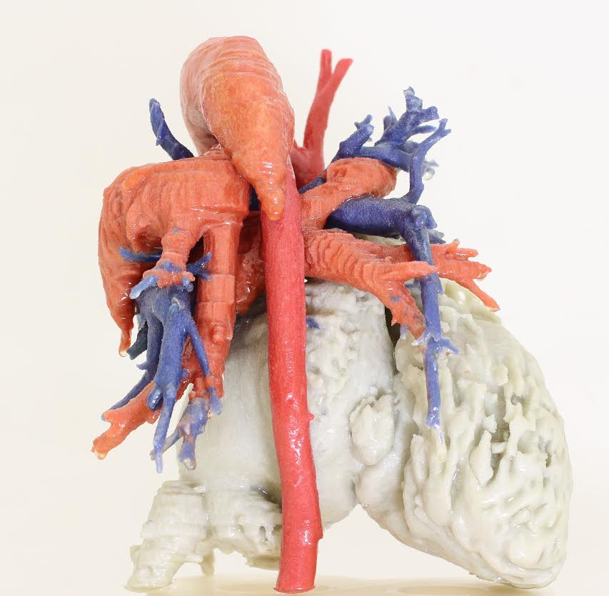
Subscribe to Our Email Newsletter
Stay up-to-date on all the latest news from the 3D printing industry and receive information and offers from third party vendors.
Print Services
Upload your 3D Models and get them printed quickly and efficiently.


You May Also Like
AM Asia Watch: China’s HeyGears Lands $44M to Expand Beyond Dental 3D Printing
Chinese 3D printing company HeyGears raised more than 300 million Yuan (roughly $44 million) in a new Series C funding round as it looks to expand beyond its industrial and...
The University of Utrecht: “3D Printing Could Change Who Gets to Become a Manufacturing Power”
For decades, manufacturing has mostly been controlled by countries with huge factories, lower labor costs, and industrial systems that took years, sometimes decades, to build. But Utrecht University human geographers...
3D Printing News Briefs, May 28, 2026: Continuous Fiber Reinforcement, Bioprinted Trachea, & More
In today’s 3D Printing News Briefs, America Makes announced the winners of its JAQS-SQ Project Call. Axtra3D is partnering with Keystone Industries to expand its dental material ecosystem, while BigRep...
Asia AM Watch: China’s SHINING 3D Restarts IPO Review Process
SHINING 3D is moving forward again with its plans to go public in China, after restarting its Beijing Stock Exchange (BSE) initial public offering (IPO) review process and filing updated...