
Researchers 3D Scan & Print Replica of 550-year Old Peruvian Baby Mummy To Better Understand Its Past

Surely, if you hopped in your handy time capsule and hightailed it back to an earlier culture to forge a discussion regarding mummies, citizens might be puzzled by our contemporary fascination–and even fear. Surely to them, it was a cultural, religious norm among the affluent, and mummies were prepared in quite a technical fashion. To find that they are something that keep children from sleeping at night still today might—or might not—be of surprise. Even if we are sometimes repelled, we just can’t look away.
Drawn in by the lore and the lure of the mysterious pyramids, their history, and the pop and horror culture we’ve created around the mummy, it’s certainly not a subject that’s dying anytime soon.
The science and study of mummies and ancient and historical artifacts is an altogether different mindset, but still undoubtedly just as mesmerizing. Recently, light has been shed on the world of mummies with the help of 3D printing technology, and this time in the instance of a 550-year-old mummy from Peru that was recovered in the desert.
Examining the ancient, mummified remains of what turned out to be a child, medical professionals at Cincinnati Children’s Hospital Medical Center were drawn into a most fascinating project due to their resources, and proximity to the Cincinnati Museum Center, where “Mummies of the World: The Exhibition” is in its final two weeks. The mummy was brought into the hospital’s radiology department so that museum experts could have help with pinpointing age, sex, and why she died. Upon some interesting findings, doctors created 3D models to help delve further into the mysteries of the particular mummy on hand.

Dr. Andrew Trout
With doctors beginning to look at and inspect the fragile, desiccated remains from within their sterile radiology department, and impart their findings, it certainly removed the mental image of the terrifying mummy we’ve all seen in the movies, bandages unraveling haphazardly as it staggers along with evil intent—for what they found beneath the remnants of mummification was the well-preserved body of a little girl thought to have been around two to three years of age at death.
“We took the same approach to evaluating these pictures of the mummy as we would to evaluating any images of any child,” said Andrew Trout, MD, a radiologist for the Department of Radiology and Medical Imaging at Cincinnati Children’s. “We are a large pediatric radiology department and within our department we are sub-specialized which takes advantage of the individual expertise of our faculty.”
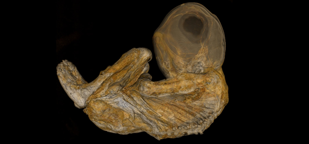
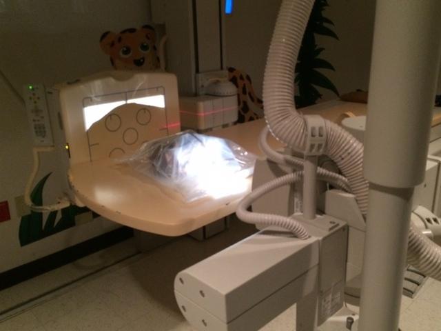
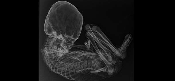
X-rays (for 2D) and CT scans (for 3D) were performed on the mummy of the little girl, and now doctors at Cincinnati Children’s have 12,000 images to pore over—taking the mummy to a whole new level of fascination—in research. Not surprisingly, doctors at the facility have been glad to volunteer their time to examine the images of the historical body, which has formally become a scientific project between the museum and the hospital.
“Dr. Alex Towbin and I are both body imagers; for this project we focused on the soft tissues to determine gender and evaluate for diseases the child might have had,” Dr. Trout continued. “Dr. Tal Laor, a musculoskeletal radiologist, was focused on the bones to try and determine age of the child and to look for finding of chronic disease or malnutrition. Dr. Jim Leach, a neuro-radiologist, was focused on the imaging of the brain and spine.”
 While unable to decide exactly what the cause of death was for the little one, the doctors did discover a hole in her spine, and that required further circumspection, which led them to thinking about using 3D printing technology to produce models that would help them research details of the X-rays and CT scans further.
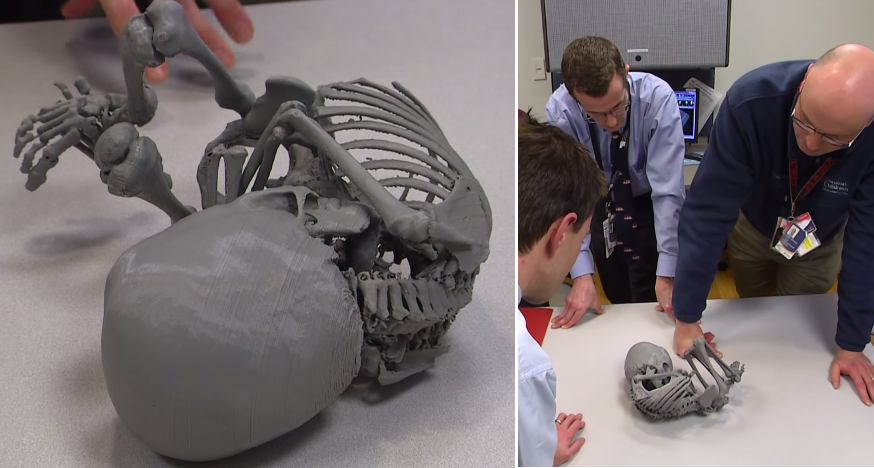
“Once I had prepared the model digitally from the scans, I printed five separate pieces and glued them together. They matched up to the exact size and position the mummy was found,” said Matt Batie, a clinical engineering specialist at Cincinnati Children’s. “I think it really helped the radiologists gather their information after being able to look closely at the model and examine it.”
Indeed, without the 3D model allowing further detailed inspection, doctors may not have been able to realize that the hole had nothing to do with her health while she was living, but rather occurred later as she was in her mummified state. While the doctors were able to find some other minor hints as to some health issues, still mysterious, they discovered she had an elongated head and weren’t sure if that was a birth defect, due to illness, or perhaps deliberate, created in a common cultural process. She also had ‘growth recovery lines,’ which could have been indication of temporary malnutrition or illness.
“Prior to the examination, we and the museum staff knew only a little about the mummy, who is believed to have been buried 400 to 500 years ago,” Dr. Trout wrote in his blog. “Unlike some mummies which are deliberately preserved and wrapped, this mummy had been preserved naturally due to the conditions where it was buried. The level of preservation of this mummy is spectacular. While the eyes are no longer present, much of the skin including ears is intact, although dried out.”
Due to the 3D models, they were able to rule out illnesses like TB, cancer, as well as death due to trauma.
“She was a relatively healthy child and we’re not sure why she died based on what we see in the images,” said Dr. Trout.
Dr. Trout and his team still have a lot of images and data to analyze, and will be spending some time in the near future doing so. “Who knows what else we will find,” he says.
Share your thoughts with us regarding this story in the 3D Printed Models of Peruvian Baby Mummy forum over at 3DPB.com. Check out more details in the video below.
Subscribe to Our Email Newsletter
Stay up-to-date on all the latest news from the 3D printing industry and receive information and offers from third party vendors.
You May Also Like
Precision at the Microscale: UK Researchers Advance Medical Devices with BMF’s 3D Printing Tech
University of Nottingham researchers are using Boston Micro Fabrication‘s (BMF) 3D printing technology to develop medical devices that improve compatibility with human tissue. Funded by a UK grant, this project...
3D Printing Webinar and Event Roundup: April 21, 2024
It’s another busy week of webinars and events, starting with Hannover Messe in Germany and continuing with Metalcasting Congress, Chinaplas, TechBlick’s Innovation Festival, and more. Stratasys continues its advanced training...
3D Printing Webinar and Event Roundup: March 17, 2024
It’s another busy week of webinars and events, including SALMED 2024 and AM Forum in Berlin. Stratasys continues its in-person training and is offering two webinars, ASTM is holding a...
3D Printed Micro Antenna is 15% Smaller and 6X Lighter
Horizon Microtechnologies has achieved success in creating a high-frequency D-Band horn antenna through micro 3D printing. However, this achievement did not rely solely on 3D printing; it involved a combination...


