Bone Regeneration Scaffolds: 3D Printing to Study Calcium Phosphate Structures


In a final degree project for Biomedical Engineering at Universitat Politecnica de Catalunya, Antonio Molina Herrero tackles the challenging topic of bone regeneration in bioprinting. Investigating 3D printing and varying material morphologies, the student researcher detailed his findings in the recently published ‘Characterization of Calcium Phosphate Structures Obtained by 3D Printing.’
In analyzing properties like the geometry of filament, porosity in structures, and surface and concavity, Herrero used micro-tomography to make comparisons with other images, calculations, and measurements—ultimately leading the researcher to understand more about deviations between nozzles and printing materials and how to use calcium phosphate better for building scaffolds in tissue engineering for bone regeneration.
“Among the synthetic bone grafts, calcium phosphate (CaPs) based ones have been widely studied. However, they do not promote osteogenesis sufficiently,” states Herrero.
“An interconnected pore network allows the vascularization of all the area and later tissue colonisation. Many studies have been carried out searching for the optimal pore size in a scaffold [1], however the conclusions are not clear and some studies contradict others; What is clear is that the pores have to be big enough to allow the vascularization ( 50 μm), but if they are larger than a certain size (500 μm) the scaffold is not acting anymore as a scaffold.”
Herrero emphasizes what he finds to be key in studying scaffolds: pore shape. Explaining that numerous methods have been used for CaP porous structures such as granulation, foaming, leaching, and freeze drying, with the advent of direct ink writing (DIW), there is even greater potential for success with such structures; however, challenges still arise for in vivo testing and further characterization.

Schema of the custom modular nozzle design. Red: Upper part of the nozzle with ¨Luer lock¨ standard connector of the top section. Grey: Lower part of the nozzle. Unscrewable to fit the custom disk. Turquoise: Disc with variety of central orifice design.
“The complex and imbricated shape of these 3D structures can be studied by advanced imaging techniques such as micro computed tomography (Micro-CT) and scanning electron microscopy (SEM). The image processing to extract quantitative information of these images is at the same time promising and challenging. These tools are increasingly used due to its immediacy and reliability. With them, morphological properties of three dimensional structures can be studied and provide morphological and structural information of a big interest for the design and characterization of new scaffolding shapes for tissue engineering,” states the author.
Samples were prepared and examined, with images and calculations being compared.
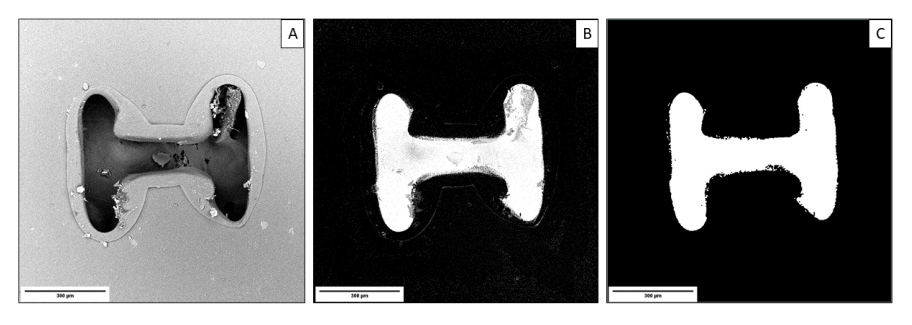
Pattern 1 image segmentation process: A) Original raw image, B) Image segmented with Ilastik and C)Image after Fiji noise reduction. 300μm scalebar.
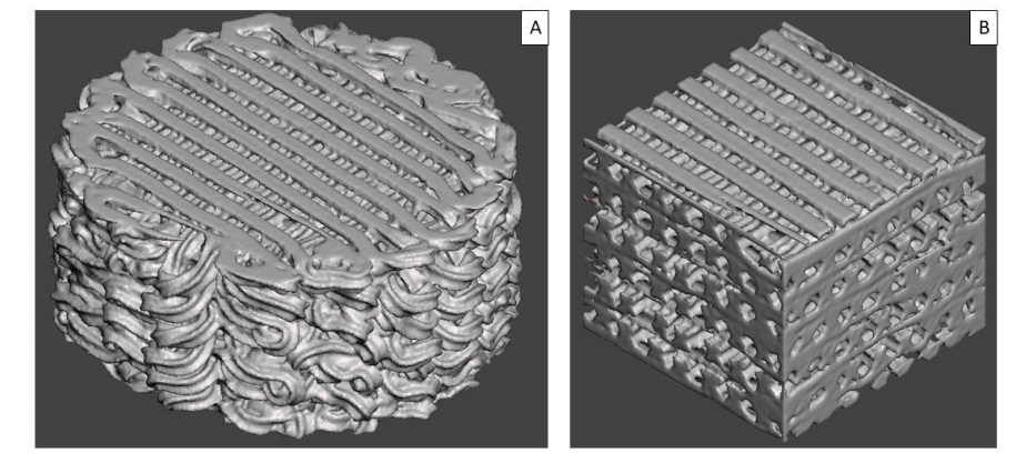
Pattern 2 before (A) and after (B) applying the VOI cropping.
Four techniques were evaluated:
- CTAn
- ImageJ
- MeshMixer
- Manual exercises
Five sample patterns were evaluated and numbered from control to 1-5.
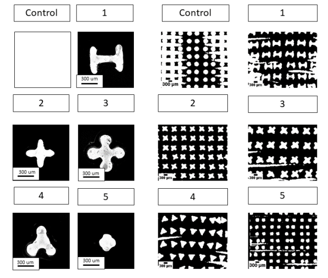
Image of the nozzles by SEM; Right: Scaffold’s sagittal view by Micro-CT.
“In the porosity results there were no large differences between the scaffolds. This it is because the scaffolds were printed in pre-setting conditions so that they would give an equivalent porosity and be able to compare the samples between them (250 μm). A good indicator about that was the pore size distribution results. The peaks in the graph represent regularity and good print quality,” concluded Herrero.
“In the specific surface area, the pattern which has the highest coefficient was the 5 and the lowest the Control. These results are coherent because the control section is a circle, so it has the least surface/area coefficient and the pattern 5; despite of being small, it was repeated very frequently, causing the sum of all the filaments to give a larger surface than the other patterns. Also, the MeshMixer method have the too much error compared to the other methods, this can be due how the plugin 3D Viewer do the meshing process from the Micro-CT scaffolds before the MeshMixer calculous, however, more trials are necessary to ensure this hypothesis.”

Porosity Percentage. Statistically significant differences indicated with different letters (p=0.05)
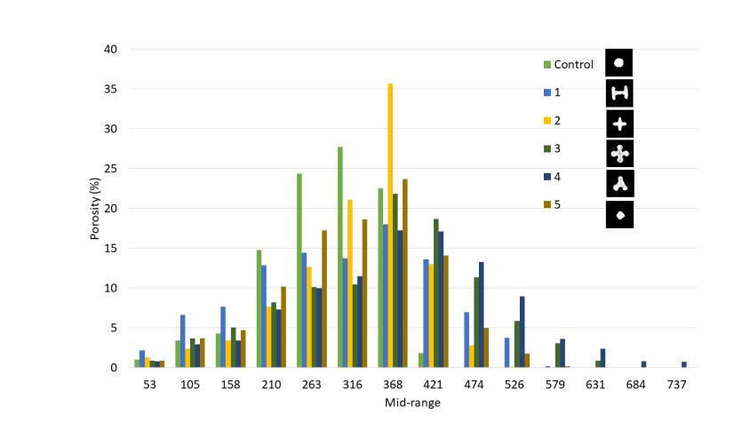
Pore size distribution.
The study of scaffolds for bone regeneration is ongoing and involved in a range of projects and experiments with the use of materials like PLA, coated nanofibers, and bioprinting with antibacterial properties.
What do you think of this news? Let us know your thoughts! Join the discussion of this and other 3D printing topics at 3DPrintBoard.com.
[Source / Images: ‘Characterization of Calcium Phosphate Structures Obtained by 3D Printing’]Subscribe to Our Email Newsletter
Stay up-to-date on all the latest news from the 3D printing industry and receive information and offers from third party vendors.
Print Services
Upload your 3D Models and get them printed quickly and efficiently.


You May Also Like
The New Dental Lab: “Three Technicians Can Handle a Hundred Arches,” Says Digital Dentistry Expert Josh Jakson
Josh Jakson’s path into digital dentistry started long before he had a job title. He grew up around it. His father, a Polish immigrant, started the family’s dental laboratory in...
AM Asia Watch: China’s HeyGears Lands $44M to Expand Beyond Dental 3D Printing
Chinese 3D printing company HeyGears raised more than 300 million Yuan (roughly $44 million) in a new Series C funding round as it looks to expand beyond its industrial and...
Asia AM Watch: China’s SHINING 3D Restarts IPO Review Process
SHINING 3D is moving forward again with its plans to go public in China, after restarting its Beijing Stock Exchange (BSE) initial public offering (IPO) review process and filing updated...
3D Printing Financials: Align’s Growth Runs on Volume
Align Technology (Nasdaq: ALGN) kicked off 2026 with steady financial results, with most of the growth coming from its core high-volume 3D printing business. The maker of Invisalign reported first-quarter...