
Slovakia: Experimenting with 3D Printed PLA Scaffolds for Bone Regeneration


Researchers from Slovakia are delving further into the uses of PLA in bioprinting, releasing their findings in the recently published, ‘3D printed Polylactid Acid based porous scaffold for bone tissue engineering: an in vitro study.’
The team created samples in the form of PLA scaffolds, assessing for both cytotoxicity and biocompatibility, with the ultimate goal of bone tissue engineering meant to assist in bone regeneration—one of the most challenging areas. Researchers have performed a wide range of different studies regarding bioprinting and bone regeneration, creating a variety of structures, scaffolds, and using different printing methods.
PLA has been used before, commonly, and while here the researchers use a commercially available type of PLA scaffolds, they separated samples into three different groups, each pre-treated with:
- Complete culture medium
- Bovine fetal serum
- Human blood
Further, the research team analyzed periosteum-derived cells in terms of cytotoxicity and biocompatibility.

Properties of used material and properties of printing process
Scaffolds were created using FFF 3D printing (bq Witbox), with scaffold samples of 10 ´ 10 ´ 4 mm and porosity of 61%. Periosteum was taken from the proximal tibia of a 55-year-old female patient who was undergoing a knee replacement surgery. Her consent was given and the Ethical Committee of The University Hospital of Louis Pasteur in Košice approved the process.
“Subsequently, cells were collected by centrifugation at 150 ´ g for 7 min and seeded 25,000 cells/cm2 in a 25 cm2 culture flask (T25) containing Alpha-modified Minimal Essential Medium (Invitrogen, GIBCO®, USA) supplemented with 10% foetal bovine serum (FBS, Invitrogen, GIBCO®, USA) and 1% ATB,” explained the researchers.
“Non-adherent cells were removed after 5 days by changing the medium. Adherent cells were cultured under standard culture conditions at 37 °C in 5% CO2 humidified atmosphere and the medium was replaced every 2–3 days. Confluent cell layers were dissociated with 0.05% Trypsin-EDTA solution (Invitrogen, GIBCO®, USA) and the number and viability of cells was assessed by TC10™ Automated Cell Counter (Bio-Rad Laboratories). Periosteum – derived osteoprogenitor cells (PDO) from the third passage (P3) – were used for the flow cytometry analysis and co-cultivation with scaffold.”
The scaffolds were sterilized, and then separated into the three groups:
“First group was incubated within human serum, second in complete medium containing Alpha- -modified minimal essential medium supplemented with 10% FBS and 1% ATB, and third in 10% FBS.”
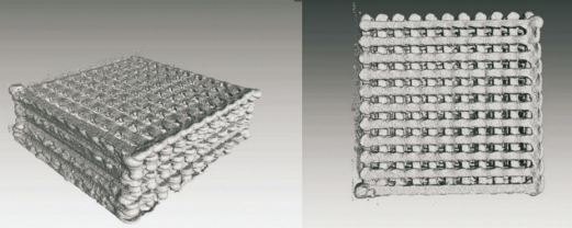
Printed scaffolds prepared by technology FFF, shown by Computed Industrial Tomography

Grafical outputs of scaffolds-internal structure of scaffold:
A) Top View (119%), B) Right view (167%)
Cells were measured four times and compared to the control group. The results yielded good biocompatibility; however, the researchers noted the best results in the scaffolds coated with human serum. This treatment also encouraged cell growth.
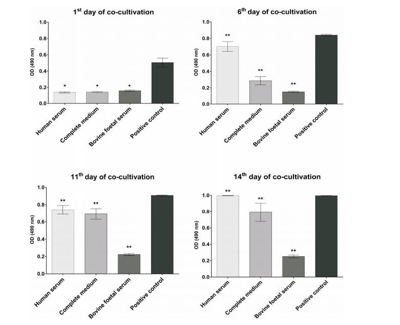
Proliferation of periosteum derived osteoprogenitors during co-cultivation with PLA scaffolds, measured after 1, 6, 11, and 14 days, respectively by CellTiter 96® AQueous One Solution Cell Proliferation Assay. Data represent mean ± SD value of four independent measures and value of p was p < 0.05 the first day of co-cultivation (*) or p < 0.01 in other days of co-culture (**)
“The distribution, adhesion and proliferation of human PDO on the native PLA scaffolds were also examined using SEM observation during two weeks of cultivation. The human PDOs showed good viability in the scaffolds, which were incubated for 48 hours in human serum, which was expressed by enhanced cellular spreading and proliferation and the pH of media, in which scaffolds and cells were co-cultivated, was 7.4 after 14 days. The pH reached the value of the one in human blood,” concluded the researchers.
“The obtained PLA porous scaffolds favored attachment periosteum derived progenitors and proliferation, furthermore, cells penetrated into the scaffold through the interstitial pores, which was meaningful for cytocompatibility evaluation. New strategies, such as poly-therapy by using scaffolds, healing promotion factors and stem cells, and finally three-dimensional printings, are in their preliminary stages, but may offer new exciting alternatives in the near future.”
What do you think of this news? Let us know your thoughts! Join the discussion of this and other 3D printing topics at 3DPrintBoard.com.
[Source / Images: ‘3D printed Polylactid Acid based porous scaffold for bone tissue engineering: an in vitro study’]Subscribe to Our Email Newsletter
Stay up-to-date on all the latest news from the 3D printing industry and receive information and offers from third party vendors.
Print Services
Upload your 3D Models and get them printed quickly and efficiently.


You May Also Like
How One Artist Is Using 3D Printing to Tell Stories About the Ocean
Artist Kimberly Callas sees something different when she looks at a 3D printer. Where others see a machine for making parts, she sees a way to tell stories about the...
Bambu Lab Wants Home 3D Printing to Feel Less Like a Workshop with PLA Pure
As desktop 3D printers become increasingly common in homes, Bambu Lab is focusing attention on something beyond print speed and hardware features. This week, the company launched a new filament,...
AM Asia Watch: China Exported 2.46 Million 3D Printers in Four Months
China’s consumer 3D printer industry seems to be reaching a new level of global dominance. According to Chinese state media outlet China Global Television Network (CGTN), China exported 2.46 million...
Bambu Launches A2L: What the New Printer Reveals About Its Strategy
Bambu Lab continues its relentless march for 3D printing domination with the launch of the A2L. The 330 × 320 × 325 mm printer will have a nozzle temperature of...


