

SLA 3D Printing Anthropomorphic Phantom Structures for Neonates


In the recently published ‘An anthropomorphic phantom representing a prematurely born neonate for digital X-ray imaging using 3D printing: Proof of concept and comparison of image quality from different systems,’ authors Nikolaus Irnstorfer, Ewald Unger, Azadeh Hojreh, and Peter Homolka explore the use of a new phantom to mimic the image of a premature newborn.
Because neonates are so miniature in comparison to adults or other children, it can be challenging to attain accurate images. As the authors point out, image processing is not generally set up to focus on this ‘special patient class,’ and the result is often that of inferior images—especially when default parameters are already in place. Neonatal patients require imaging at low doses, which necessitates high-performing equipment.
Most other structures fall short too, including using animal models, where ‘interobserver variability in judgment of image’ was high. The authors realize these problems could be overcome if identical patient equivalent radiation patterns were possible allowing for:
- Direct side-by-side comparison between different detector systems
- Different image processing algorithms
- Different acquisition settings
With image acquisition in place correctly, dose levels could be adjusted downward. The authors point out that this is a common approach and one that demonstrates the potential for advanced image processing.
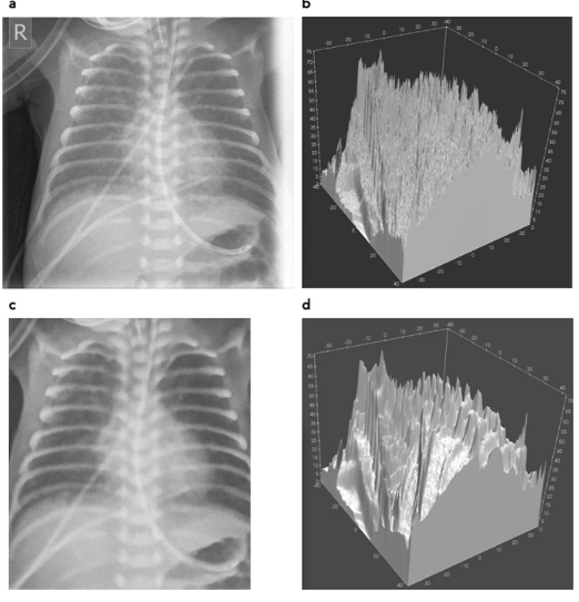
Patient image used as printing template, and 3D visualization of the respective phantom STL models before and after de-noising. (a) Diagnostic patient image without noise reduction, and (c) after cropping and noise reduction. Corresponding phantom STL models are shown in (b,d).

Phantoms printed with FDM (left) and SLA (right image) technology, respectively (a), and corresponding x-ray images (b: FDM, and c: SLA). Better reproduction of sharp structures and fine details with SLA is evident.
SLA 3D printing stood out as the superior method for re-creating small details in image processing.
“Better reproduction of details can be seen in the SLA printout e.g. in the catheter walls in the upper left corner of the image, and in the reproduction of the spinous processes. Therefore, the SLA print was chosen for further evaluation and subsequent use as phantom to be used for optimization exercises in clinical systems,” stated the authors.
Phantom images from other systems were compared, with the differences helping radiologists understand more about optimization.
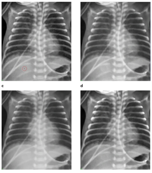
Phantom images from different systems normalized to identical level and latitude. (a) Agfa needle type storage phosphor (reference system), (b) system 2, (c) system 3, and (d) system 4. In (a) ROI in the homogeneous disk in the liver region used for noise measurements is marked (red circle).
“Since patient images taken at extremely low doses exhibit high quantum noise, however, noise removal has to be applied. Different algorithms also used in clinical image processing have been developed to best preserve structure. Denoising, even if structure preserving, always degrades detail,” concluded the researchers. “In this work black-box algorithms implemented into a professional software package optimized for digital photography have been used. Lung structure and bone contours are affected by these algorithms. Also, visual inspection of critical structures as opposed to quantitative measures were applied to define the level of noise reduction to be used in the printing template. However, deciding on the level of de-noising remains subjective because limited by the accepted remaining noise, and acceptable loss of detail.
“…future development of these phantoms should consider integration of some kind of realistic fine-structure in the lungs mimicking also spectral properties of the patient lung.”
3D printing has been associated with improvements in the use of phantoms for image processing, from those created for cardiac patients, to assistance in treatment for cancer patients, uses for determining radiation dosages, and more. What do you think of this news? Let us know your thoughts! Join the discussion of this and other 3D printing topics at 3DPrintBoard.com.
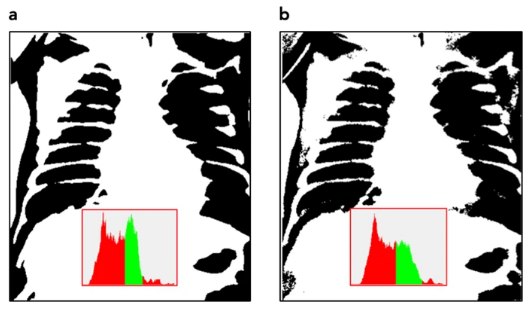
Thresholded binary images indicating pixel intensities corresponding to lung tissue (black). Threshold settings correspond to green part in the histograms. (a) de-noised patient image, (b) phantom image.
Subscribe to Our Email Newsletter
Stay up-to-date on all the latest news from the 3D printing industry and receive information and offers from third party vendors.
Print Services
Upload your 3D Models and get them printed quickly and efficiently.


You May Also Like
Divergent Declares that German 3D Printers are Superior, And Plans Massive LPBF Expansion
Divergent has announced a new version of its Laser Powder Bed Fusion (LPBF) printer and a new site. The company aims to do nothing short of “further accelerating its mission...
Incodema3D Buys 14 Metal EOS Systems, Now One of the World’s Largest Metal 3D Printer Operators
Recently, a majority stake of 3D printing service bureau Incodema3D was purchased by AFM Capital. Under new ownership, the Freeville, New York company is now using its cash-rich parent for...
CEO Yoav Zeif on Why Stratasys’ Markforged Acquisition Is Really a Bet on Industrialization
When Stratasys announced plans to acquire Markforged, the immediate focus was on the deal. Markforged is one of the most recognizable names in additive manufacturing (AM), known for its continuous...
3D Printing & the Autonomous Era: Defense Tech’s Latest Mutation
When we last checked in on the broad defense tech landscape and the role of the additive manufacturing (AM) industry in that environment, it became clear that the connecting thread...


