
A Window Into the Brain: Transparent 3D Printed Mouse Skulls for Alzheimer’s Research


There’s a long way to go to understanding the brain. So many questions remain unanswered, yet so much research is being done, like the former U.S. administration’s ambitious project, which Barack Obama called the BRAIN initiave, attempting to map the activity of every single neuron to understand brain circuits. Or the European Union’s Human Brain Project, a conjoint research study that allows scientists to advance their knowledge of neuroscience. Researchers have tried to tackle the underlying causes of the most common illnesses that alter the brain, yet Alzheimer’s, a disease that affects over 44 million people worldwide with a global cost estimated at $605 billion, is expected to reach as many as 135 million people by 2050, while another 10 million people around the globe already suffer from Parkinson’s. Labs and pharmaceutical companies are developing ways to help people via inhibitors and stem cell therapy, but haven’t come to a definitive solution, something most specialists think could take years.
Using 3D printing researchers have developed a technique that could aid science’s path to a better understanding of the brain. Last April, a team of researchers at the School of Medicine’s Department of Neuroscience at the University of Minnesota developed a unique 3D printed transparent skull implant for mice that provides an opportunity to watch activity of the entire brain surface in real time. It is like having a window to the brain and being able to see how the fully functional organ really works, what stimulates, harms and overall degenerates it. The innovative device called See-Shell could provide new insight for human brain conditions such as concussions, Alzheimer’s and Parkinson’s disease, and will soon be sold commercially.
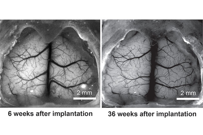
The whole cortical surface of the mouse at six weeks and 36 weeks after implantation of the 3D printed See-Shell.
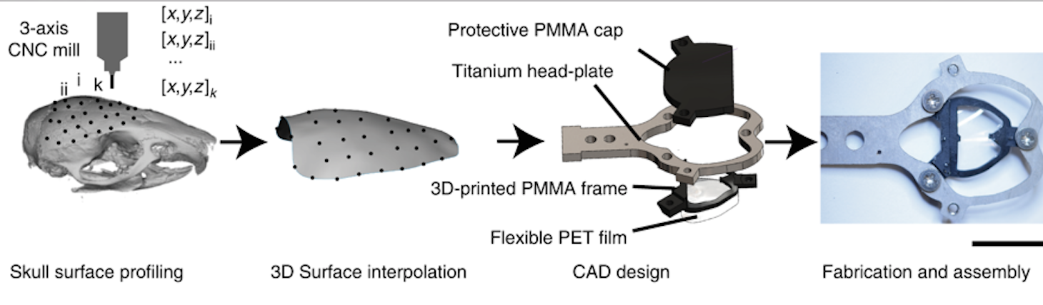
The digitally designed, morphologically realistic, transparent polymer engineered See-Shell skulls allow researchers to see the way mice brains function during long periods of time, that is 300 days or more, as well as using chronic imaging, which provides an opportunity to study brain development, plasticity, learning, disease processes and evaluation of new therapies. To actually make the skull, the researchers first digitally scanned the surface from a single mouse skull, which served as a template to design the generalized transparent skulls using CAD software. The frame was then 3D printed out of polymethylmethacrylate (PMMA) onto which a thin, flexible and transparent polyethylene terephthalate (PET) film was bonded – PET was chosen for its transparent element as this polymer has excellent optical properties and is biocompatible. The 3D printed frame also incorporates screw holes for fastening a custom-designed titanium head-plate for head-fixing the animal during experiments. Once the See-Shell was finished, a part of the mouse’s skull was carefully removed through surgery and replaced with the 3D printed implant. The researchers used a robot with surface profiling capacities to guide a computer numerical controlled (CNC) mill to perform the craniotomy.
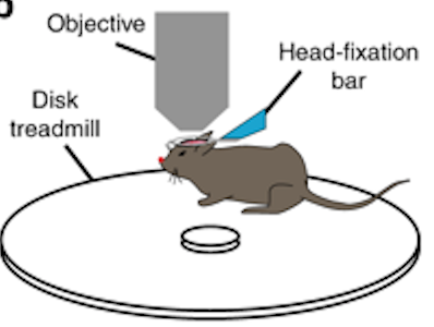
“What we are trying to do is to see if we can visualize and interact with large parts of the mouse brain surface, called the cortex, over long periods of time. This technology allows us to see most of the cortex in action with unprecedented control and precision while stimulating certain parts of the brain.This will give us new information about how the human brain works,” says Suhasa Kodandaramaiah, co-author of the study and University of Minnesota Benjamin Mayhugh Assistant Professor of Mechanical Engineering.
With an overall surgery success rate on mice of 93.5%, the lab creatures implanted with See-Shells readily performed a variety of behaviors including walking and grooming during head-fixation. According to the team, one of their first studies using the See-Shell device examines how mild concussions in one part of the brain affect other parts of the brain as it reorganizes structurally and functionally. According to Kodandaramaiah, “mouse brains are very similar in many respects to human brains”, and this device opens the door for similar research on mice looking at degenerative brain diseases that affect humans like Alzheimer’s or Parkinson’s disease.
According to the research paper, entitled “Cortex-wide neural interfacing via transparent polymer skulls,” which can be found in the journal Nature Communications, several aspects of the design and fabrication of the See-Shells are widely adoptable and highly flexible. See-Shells can be fabricated using desktop tools and are inexpensive, under $20 each, and once the individual components are fabricated, the implant can be assembled in less than 15 minutes. Therefore, the researchers consider this a tool that can be readily adopted by most laboratories. Although the cranial implants were developed for the dorsal cerebral cortex with its fairly regular convex surface, the design can be modified for a variety of skull morphologies. As reported by the scientists in charge of the project, future versions of the 3D printed skull can be designed to cover not only the dorsal cerebral cortex but also other regions including the olfactory bulb, cerebellum, and more lateral cortical regions such as the auditory cortex. The CAD files used for 3D printing of the frame are also available for download.
The 3D printed transparent skull implant for lab mice
One of the most important aspects of the development is its see-through structure, which allows for multi-scale imaging over long periods of time in the same animal and with sub cellular resolution. This will provide new insights, such as physical structure and neural state. Additionally, the technology allows researchers to see global changes for the first time at an unprecedented time scale and detail, such as changes in brightness of the mouse’s brain which correspond to waxing and waning of neural activity, while subtle flashes are interpreted as periods when the whole brain suddenly becomes active. It might take some time before specialists understand what all this means, still, the application of 3D technology has definitely opened a new way to understand how to help the brain recover from illnesses or even prevent them.
Timothy J. Ebner, co-author of the study and Head of the Department of Neuroscience at the University of Minnesota, claimed these studies “couldn’t be done in humans”, but they are extremely important to the understanding of how the brain works “so we can improve treatments for people who experience brain injuries or diseases.” The lab mice used during the research were allowed to recover from surgery for at least seven days before imaging experiments were attempted, and after implantation, mice were allowed to recover on a heating pad until ambulatory and then returned to a clean home cage. They were all administered opioid derivatives post-operatively on the day of the surgery as well as during the three succeeding days to assist with full recovery.
The 3D printed See-Shell device created at the University of Minnesota
This is not the first time that University of Minnesota researchers have targeted Alzheimer’s: during the last few years they have teamed up with startup RetiSpec to commercialize technology for early detection of Alzheimer’s, and identified a target that could be used to develop new treatments to restore communication between neurons within the brain and applied a retinal scan technique, developed by researchers in the University’s Center for Drug Design (CDD), to diagnose early-stage Alzheimer’s disease by looking into a person’s eye.
Scientists and researchers have been working during the last few years on developing new treatments for brain injury and illness using 3D printing. The technology can go a long way to understanding basic brain activity and aiding in the development of better solutions for patients. Two years ago scientists at the University of Wollongong-based ARC Centre of Excellence for Electromaterials Science (ACES) began bioprinting tissue from human-induced pluripotent stem cells (iPSCs), which are stem cells that have the capability of differentiating into any type of adult cell, including brain cells, and in the future, the team plans to 3D print brain cells that produce dopamine as a possible treatment for Parkinson’s disease. While a team of Harvard University scientists even ran a series of experiments using a 3D printing technique that mimicked the growth and development of a human brain; and Oklahoma State University researchers are investigating the use of new materials and structures for successful neural tissue bioprinting.
The research was funded primarily by the National Institutes of Health (NIH) with additional support from Minnesota’s Discovery, Research, and InnoVation Economy (MnDRIVE) funding from the State of Minnesota. While the imaging research was made possible by the cutting-edge imaging infrastructure at the University Imaging Center. While there is no cure for Alzheimer’s disease or even a way to stop its neuron-degenerative progression, the researchers are opening a whole new world of possibilities through their novel design, another way 3D printing is proving to be an ideal bespoke solution to some age-old diseases, and could eventually help individuals living with the disease improve their quality of life.
Subscribe to Our Email Newsletter
Stay up-to-date on all the latest news from the 3D printing industry and receive information and offers from third party vendors.
Print Services
Upload your 3D Models and get them printed quickly and efficiently.


You May Also Like
Australia’s AMCRC Funds Titanium 3D Printing R&D
In terms of the global economy’s presently existing state, there is no realistic path to economic resilience that doesn’t start with critical minerals security. This is a problem for pretty...
3DPOD 305: Automating AM with Grenzebach’s Oliver Elbert
Oliver Elbert‘s over ten years in additive manufacturing have been spent automating LPBF. For large, high-volume, or critical parts, Grenzebach has provided custom automation solutions. Depowdering, powder handling, sieving, heat...
AMPulse Asia: Chinese IPOs, Defense Deals, and Dental 3D Printing Lead APAC Roundup
The second half of June brought a wave of additive manufacturing activity across China, Japan, South Korea, India, and Australia. From Chinese IPOs and funding rounds to defense, aerospace, construction,...
Austal, Curtin University and AMCRC Work on R&D Together
Australia’s Additive Manufacturing Cooperative Research Centre (AMCRC) works with 70 industry partners to deliver collaborative R&D projects. They also work on workforce development and technology transfer. It’s kind of analogous...


