
3D Printing Helps Researchers Discover Why the Human Brain Has a Folded Structure

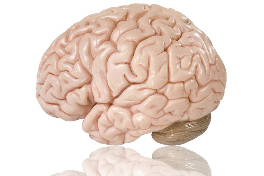 Despite its importance, the human brain still remains a huge mystery to most of medical science. While many researchers believe that the human brain has a folded structure in order to increase the amount of surface area that can fit into the smaller volume available inside of the human skull, the truth is that medical science doesn’t really know why that structure exists. While that lack of knowledge isn’t for want of trying, the medical ethics involved with running experiments on the human brain make it difficult to conduct substantive research using live models.
The brain’s growth process, which is known as gyrification, has generally been believed to be a biological response to the need for human brains to maximize the number of cortical neurons while minimizing the distance between them. However a team of researchers ran a series of experiments using a 3D printing technique that mimicked the growth and development of a human brain, which typically begins at about the 23rd week of gestation and continues until well into adulthood. The researchers believe that they have proven that the folded structure is simply a physical growth process and not a matter of biology.
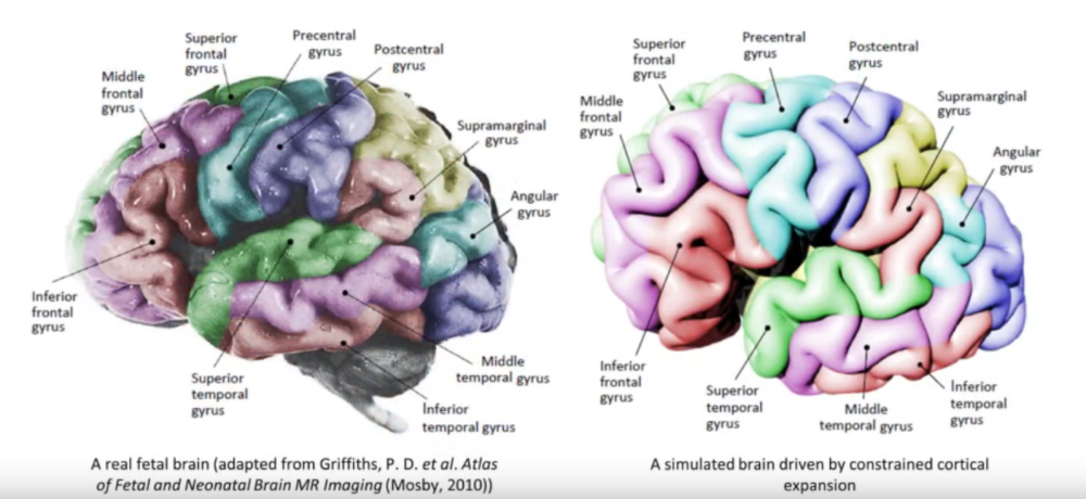
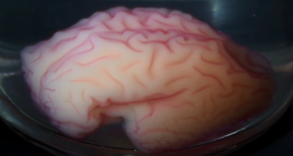
Comparisons between a real brain and a brain generated using a simulation.
The results of the team’s experiments were published in Nature Physics this month as a paper called “On the Growth and Form of Cortical Convolutions”. The team of Harvard University scientists based their research on a little known model of brain development that was created more than 40 years ago. The model suggested that the shape and structure was simply the result of a growth process and not directly tied to any biological the chemical directives from the brain. The researchers’ experiments with a 3D printed simulation of the brain seem to prove that the folds are actually the result of mechanical compression forces in response to the brain’s rate of growth. The beneficial side effects of the placement of cortical neurons in the brain seems to be a response to the brain’s growth process, not an active motivator for the brain’s growth process.
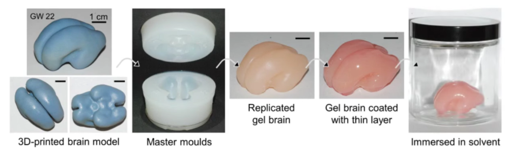
The 3D printed foetal brain.
In order to run their simulation of brain development the team 3D printed a model of a fetal brain based on MRI data using several different layers of soft gel materials. The layers of gel were designed to swell and expand at different rates when placed in a specialized liquid solvent. The process was designed to simulate the growth and development of the brain’s gyri and sulci, the areas of folded tissue on the surface of the brain. The 3D printed model of the brain accurately developed folds on its surface in the same patterns as those observed in real human brain development as the outer layer compressed in on itself in response to the expansion of the inner layers of gel.
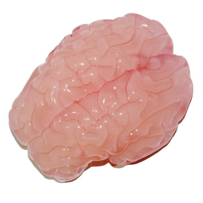
The 3D printed brain after being immersed in the solvent.
“Starting with the same initial geometry, we also build numerical simulations of the brain modelled as a soft tissue with a growing cortex, and show that this also produces the characteristic patterns of convolutions over a realistic developmental course. All together, our results show that although many molecular determinants control the tangential expansion of the cortex, the size, shape, placement and orientation of the folds arise through iterations and variations of an elementary mechanical instability modulated by early foetal brain geometry,” wrote the study authors Tuomas Tallinen, Jun Young Chung, François Rousseau, Nadine Girard, Julien Lefèvre and L. Mahadevan.
The research could lead to more accurate methods of diagnosing and treating many neurological disorders that have been linked to the malformation of the brain, as well as brain cell migration and the thickness of the cortex. While this simulation of the development of the brain surface will provide a useful template for future studies, the process of recreating the very complicated growth process of a real human brain, and how that relates to how it functions, is quite challenging. This model is limited only to predicting the behavior of simple, basic brain structures at the very onset of the folding process.
The 3D printed brain during the formation of the simulated brain folds.
“Their simulations explain why folding always begins in weakly curved regions and why gyri and sulci align perpendicular to the direction of maximum compressive stress. Experiments with swelling brain models provide the essential missing link between modelling, experiment and simulation. However, a few limitations remain: the model is beautiful and simple, but it is limited to the initial folding of idealised structures; the experiment is useful for exploring instabilities beyond the onset of folding, but it is limited to moderate changes in volume,” explained the Departments of Mechanical Engineering and Bioengineering at Stanford University in California’s Ellen Kuhl in an accompanying paper that she authored, also published by Nature Physics.
You can see a video overview of the experiment here:
The experiment is potentially a starting point for new lines of research that could help doctors and neuroscientists develop new understanding of the human brain which could lead to several medical advancements. Specifically, being able to link the rate of brain growth to neurological development could help scientists trace individual brain functions back to the folding of the brain surface. Because the process is mechanical, not biological, it could accurately determine how and when something goes wrong. This could lead to the ability to identify surface markers that may lead to the early diagnosis of autism, schizophrenia or Alzheimer’s disease. Early detection could also lead to the development of more effective treatment options and the cultivation of new preventative measures. Discuss this amazing new research in the 3D Printed Brain Model forum over at 3DPB.com.
Subscribe to Our Email Newsletter
Stay up-to-date on all the latest news from the 3D printing industry and receive information and offers from third party vendors.
Print Services
Upload your 3D Models and get them printed quickly and efficiently.


You May Also Like
Reinventing Reindustrialization: Why NAVWAR Project Manager Spencer Koroly Invented a Made-in-America 3D Printer
It has become virtually impossible to regularly follow additive manufacturing (AM) industry news and not stumble across the term “defense industrial base” (DIB), a concept encompassing all the many diverse...
Inside The Barnes Global Advisors’ Vision for a Stronger AM Ecosystem
As additive manufacturing (AM) continues to revolutionize the industrial landscape, Pittsburgh-based consultancy The Barnes Global Advisors (TBGA) is helping shape what that future looks like. As the largest independent AM...
Ruggedized: How USMC Innovation Officer Matt Pine Navigates 3D Printing in the Military
Disclaimer: Matt Pine’s views are not the views of the Department of Defense nor the U.S. Marine Corps Throughout this decade thus far, the military’s adoption of additive manufacturing (AM)...
U.S. Congress Calls Out 3D Printing in Proposal for Commercial Reserve Manufacturing Network
Last week, the U.S. House of Representatives’ Appropriations Committee moved the FY 2026 defense bill forward to the House floor. Included in the legislation is a $131 million proposal for...