
University of Pittsburgh: 3D Printing Mammary Glands to Study Premalignant Disease


 Recently, Adrian V. Lee of University of Pittsburgh prepared a study for the U.S. Army Medical Research and Materiel Command regarding 3D printed medical models and breast cancer research. His findings are outlined in ‘A 3D Bioprinted Model for the Study of Premalignant Disease,’ published by the Defense Technical Information Center.
In hypothesizing that in vitro 3D bioprinted models of premalignant breast cells could help identify markers for low-risk premalignant disease, the research team was comprised of the following specialists as ultimately, they endeavored to 3D print mammary glands:
- Surgical oncologist
- Mammary gland biologist
- Biomedical engineer
- Cancer biologist
Goals were designated for each year of the study, and completed as follows:
- Year 1 – Quantify mammary gland development and find strain dependent differences.
- Year 2 – Keep characterizing development of the mammary glands.
- Year 3 – Study growth patterns of breast cells in vitro.
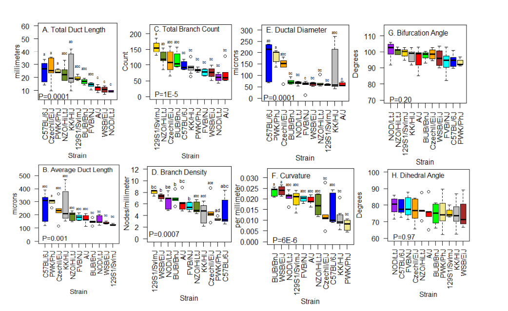
Genetic Background influences overall mammary ductal geometry. Three-dimensional reconstructions were prepared from E-cadherin-stained mammary whole mounts. Tissues were collected from females of 11 different inbred strains at post-natal day 17 of age. Shown are (A) total duct length, (B) average duct length, (C) total branch count, (D) branch density, (E) ductal diameter, (F) curvature, (G) bifurcation angle, and (H) dihedral angle. Each box represents the data for 3 to 7 animals. Statistical significance was set at a=0.05. Boxes are ordered by strain median. Boxes with similar superscripts are similar (P>0.05) by Tukey’s HSD.
Mice were used extensively in the study too, with their ages corresponding to the onset of human puberty, and mammary glands not yet affected by estrus. With breeding pairs, the researchers were able to use mammary tissue for 3D imaging that could then be sent to University of Pittsburgh for 3D printing.
“This work represented the first 3D comparison of ductal architecture and patterning in inbred mouse strains of different genetic backgrounds,” states Lee. “The hypothesis for the study was that ductal patterning, and the implementation of stereotypical branching behaviors during early post-natal development differs with genetic background.”
The research team noted differences in:
- Total duct length
- Average duct length
- Total branch count
- Branch density
- Ductal segment diameter
- Curvature
As work progressed, the researchers began bioprinting directly with collagen Type 1 and additional ECM protein hydrogels:
“The mammary duct model we developed represents a world-first level of complexity generated using a bioprinter with multiple ECM and hydrogel components.”
In the next phase, the researchers used progenitor cells to 3D print the mammary ductal structure. Beyond that, they completed further imaging and assessing of the 3D progenitor cell (still ongoing).

Delamination of collagen is consistent across constructs seeded and cultured for a week. Constructs shown on the left were cultured with MCF7 cells, and constructs on the left were cultured with MCF10a cells. In all constructs, a gap of cells was visible around the top rim of collagen, indicating that the collagen was originally there but pulled away under the action of cells. Otherwise, we would expect to see cells scattered around the edge of the rim on all regions of top-side alginate. Scale bars are 1 mm.
Challenges occurred as they noticed a loss of structure in the models as cells began to affect the collagen detrimentally. All the bioprinted structures experienced delamination, no matter what types of cellular foundation they possessed. Growth was interrupted and characterization overall ‘inhibited.’ They redesigned the structure for better growth and quality in their research overall.
This new system, printed entirely in collagen type I, was fabricated with an inner diameter of 1.4 mm, matching the average breast. The team has now 3D printed over 25 of the bioprinted models successfully.

The redesigned mammary duct construct showing the simplified tube design and perfusion validation. (A) Schematic of the construct. (B and C) Photographs of the construct printed entirely from collagen type I using the FRESH 3D bioprinting method. (D) Perfusion studies using our bioreactor platform to perfuse fluorescent Dextran of various molecular weights through the lumen and tracking diffusion through the tube wall. (E) Quantification of the perfusion and permeability studies in (D), showing as expected that diffusion through the wall depends on molecular weight, but also that there are no large defects.
“In year 1 we encountered minor difficulties such as reduced fecundity in some mouse strains, but we continued these studies in year 2 and complete the mammary ductal development studies as noted above. During year 2, we had to change the design of the 3D ductal microenvironment, as the method we developed at the end of year 1 showed uneven plating of cells and delamination of collagen,” concluded the researchers. “We re-engineered the 3D ductal environment to allow perfusion and easier plating of cells. Preliminary results show that this model has better cell plating and cell survive when toxins are removed by perfusion. In year 3 (NCE) we will now study growth of cells in this microenvironment.”
3D printed medical models have made significant impacts in the past few years especially as more scientific facilities and hospitals have begun building labs onsite. From using them as guides to rebuild areas as complex as an eye socket to training medical students and even bioprinting brain tumors to learn more, models, surgical guides, implants, and devices, and changing the face of medicine today—and the lives of patients around the globe. Find out more about how 3D printed models may help in the study of breast cancer here. What do you think of this news? Let us know your thoughts! Join the discussion of this and other 3D printing topics at 3DPrintBoard.com.
[Source / Images: A 3D Bioprinted Model for the Study of Premalignant Disease]Subscribe to Our Email Newsletter
Stay up-to-date on all the latest news from the 3D printing industry and receive information and offers from third party vendors.
Print Services
Upload your 3D Models and get them printed quickly and efficiently.


You May Also Like
APAC’s 3D Printing Capital Wave Is Bigger Than Venture Funding
By the usual measure, a tally of funding rounds, APAC’s additive manufacturing market had a quiet second quarter. The capital that has actually closed across the region comes to about...
HP Stock Jumps on 3D Printing Buzz
HP (NYSE: HPQ) had its best day in over a year this week, with shares jumping more than 7% on Tuesday. Interestingly, the move was quickly tied to 3D printing,...
3D Printing News Briefs, April 8, 2026: LiDAR Scanning, Vapor Smoothing, FDM Optimization, & More
We’ll kick off today’s 3D Printing News Briefs with some 3D scanning news from Artec 3D, and then move on to new America Makes Project Calls. Then, Raise3D and AMT...
3D Printing Market Hits $16B in 2025 as Growth Picks Up Again
The global 3D printing market reached $16 billion in 2025, growing just over 10% year over year, according to new data from Additive Manufacturing Research (AM Research). After a slower...


