Bioprinting 101 – Part 12, Phantoms

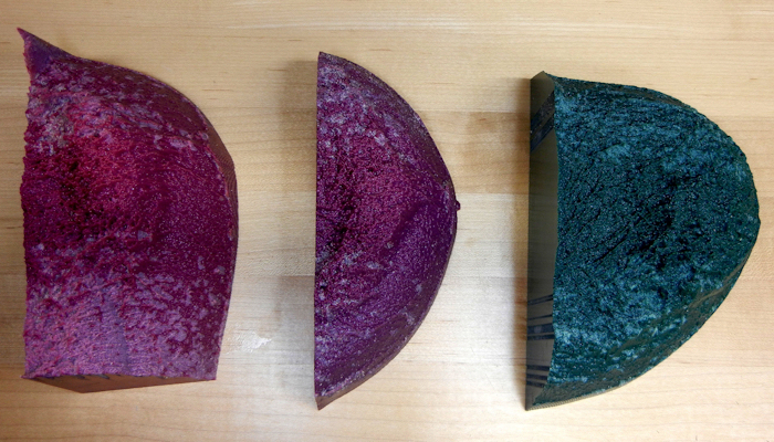
Within the bioprinting area, a phantom refers to a model of the body or of a specific part thereof. Typically we are using replicas of an organ to model it, learn and to experiment with it outside of the body. One may think in terms of a heart. If a heart has a valve that may not be in the best geometric alignment, we can replicate the heart within a human and use this phantom to experiment surgically outside of the patient’s body to ensure better results for an actual procedure. With 3D bioprinters, we are mimicking specific organs that are within an individual human. There lies a lot of importance to this mimicking. This can be used to help students within medical schools, as well as practitioners within their actual fields. Complex geometries of organs are specific to different people. Having insight into various differences from person to person through phantoms allows us to learn more in-depth about the human body and its parts. Phantoms are not for use within a patient. They are used for practice outside of a patient typically.
How are these phantoms created then? They are typically made through the use of imaging technologies that are present within the medical field. These technologies include the following and are not limited to:
- CT
- MRI
- PET
- SPECT
- Ultrasound
- Elastography
We will briefly talk about these methods, but will outline them further within our series on bioprinting.
Phantom Image vs Real Organ image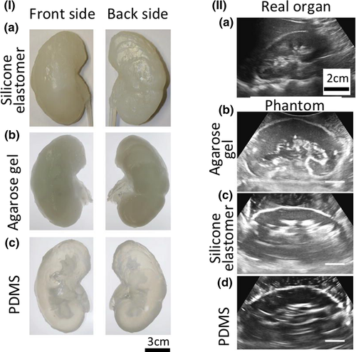
A CT scan, also known as computed tomography scan, and formerly known as a computerized axial tomography scan or CAT scan, makes use of computer-processed combinations of many X-ray measurements taken from different angles to produce cross-sectional (tomographic) images (virtual “slices”) of specific areas of a scanned object, allowing the user to see inside the object without cutting.
An MRI is a medical imaging technique used in radiology to form pictures of the anatomy and the physiological processes of the body in both health and disease. MRI scanners use strong magnetic fields, magnetic field gradients, and radio waves to generate images of the organs in the body.
Positron-emission tomography (PET) is a nuclear medicine functional imaging technique that is used to observe metabolic processes in the body as an aid to the diagnosis of disease. The system detects pairs of gamma rays emitted indirectly by a positron-emitting radioligand, most commonly fluorine-18, which is introduced into the body on a biologically active molecule called a radioactive tracer.
Single-photon emission computed tomography (SPECT, or less commonly, SPET) is a nuclear medicine tomographic imaging technique using gamma rays. It is very similar to conventional nuclear medicine planar imaging using a gamma camera (that is, scintigraphy) but is able to provide true 3D information. This information is typically presented as cross-sectional slices through the patient, but can be freely reformatted or manipulated as required.
Medical ultrasound (also known as diagnostic sonography or ultrasonography) is a diagnostic imaging technique based on the application of ultrasound. It is used to create an image of internal body structures such as tendons, muscles, joints, blood vessels, and internal organs. Its aim is often to find a source of a disease or to exclude pathology.
Elastography is a medical imaging modality that maps the elastic properties and stiffness of soft tissue. The main idea is that whether the tissue is hard or soft will give diagnostic information about the presence or status of the disease. For example, cancerous tumors will often be harder than the surrounding tissue, and diseased livers are stiffer than healthy ones.
After a scan, typically a phantom is used for assessing the status of an organ within the body without the need for invasive operations to be done. Being able to replicate an organ in such a manner is definitely beneficial. It allows doctors to have less of a need to cut open a patient to dissect the problems of their specific organ. It is a good initial check and it is good for preventing unnecessary operation. It also leads to an easier diagnosis of problems within an organ. One can likely use a phantom for transplant purposes, but this is not necessarily feasible yet. The importance of resolution and detail is too high to reliably replicate an organ for transplant use at the moment.
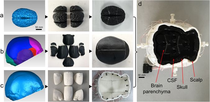
In general, images taken with the technology listed above are in 2D form. In order to have a representation of an object as a whole, we need to see it within a 3D way. This requires us leveraging multiple images within the 2D form in a process called image stitching. Image stitching or photo stitching is the process of combining multiple photographic images with overlapping fields of view to produce a segmented panorama or high-resolution image. Commonly performed through the use of computer software, most approaches to image stitching require nearly exact overlaps between images and identical exposures to produce seamless results. To create a 3D form through the use of 2D images, we must rotate the images through computer software and then optimize algorithms to stitch a 3D form based on rotational image data. Software such as Materialise Mimics is used to generate STL data from the Dicom files that are the output of CT or MRI scans. The STL can then be 3D printed. Nowadays an emerging trend is to have phantoms made on desktop 3D printers which makes them more accessible and less expensive. SLA printers such as the Formlabs or FDM machines such as inexpensive Prusa’s have been used to generate phantoms that cost only a few dollars. Industrial 3D printers such as the Stratasys J750 have also been used to give gradient parts soft areas for example while other prints use industrial stereolithography or DLP machines to make for highly accurate parts.
The importance of imaging techniques and creating phantoms lies within the precision of a 3D image. We want to highest quality resolution. This allows for reconstruction and building of a phantom that best mimics the organs that may lie within a human’s specific body. We will analyze this importance of precision within future articles that outline these different techniques and their relation to bioprinting as a whole.
Subscribe to Our Email Newsletter
Stay up-to-date on all the latest news from the 3D printing industry and receive information and offers from third party vendors.
You May Also Like
NSF Awards Kentucky $1M for Advanced Manufacturing
The National Science Foundation has awarded a $1 million grant to the University of Louisville for the Advancing Manufacturing and Building Construction Technologies (NSF AMT) project. This initiative is part...
3D Printing News Briefs, May 11, 2024: 3D Printed Stent, Tower, Sculptures, & More
We’re starting off with medical research in today’s 3D Printing News Briefs, as researchers in Korea used CT images and 3D printing to fabricate an educational simulator for a mastoidectomy....
3D Printing Unpeeled: Wind Turbines, Probiotics and Lenses
TPI Composites, ORNL and Ingersoll Rand are working to make wind turbine tooling segments that can be 18.3 meters long. These elements also include resistive wires that help keep the...
Tethon 3D Releases Cost-effective Bioprinter
Tethon 3D, known for its ceramic-loaded DLP materials, custom resins, and DLP 3D printers, has recently released a bioprinter. Vat polymerization printers like DLP systems have been widely used by...