Holding Their 3D Printed Heart is Helping Patients and Their Doctors

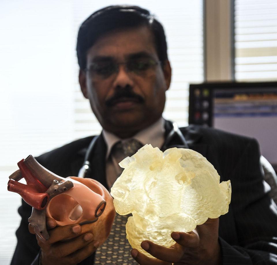
Dr. Joseph Vettukattil comparing an education heart model and Borgman’s 3D printed heart.
The practice of 3D printing replica organs to prepare for long, complicated surgeries is starting to become a regular part of the operative pre-planning process. Being able to hold a physical replica of the exact organ that they will be operating on is becoming an invaluable tool for surgeons. Often it can shave hours off of the procedure time, not to mention giving doctors the ability to anticipate any potential difficulties they may encounter during the surgery, and plan ways to avoid them. But the practice is also having an unexpected benefit in offering the actual patient the opportunity to see the model and give them the ability to understand and visualize what exactly is being done to them.
When Nicholas Borgman and his twin sister Alyssa were born in 1989, their parents Vicki and Jerry received some alarming news. While Alyssa was in perfect health, Nicholas was born with a serious heart defect called a pulmonary atresia with an intact septum. The defect in his pulmonary valve hindered the flow of blood from Nicholas’ heart to his lungs, and he needed to have heart surgery immediately. His first surgery was at only six days old, his second at seven months old, and his final was at two years old. Thanks to these life-saving surgeries Nicholas went on to lead a healthy and active childhood, but unfortunately that wasn’t the end of his heart problems.

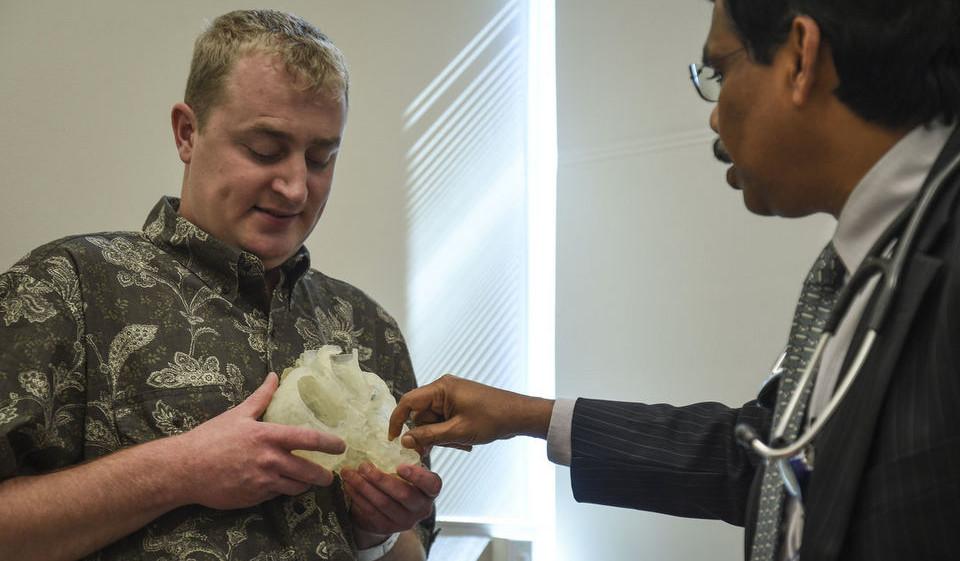
Dr. Vettukattil explaining the heart surgery to Nicholas Borgman and his family.
At 25, Borgman saw his doctor for a routine checkup and discovered that his heart had an enlarged right chamber caused by high pressure building up in the right side of his heart. The left side of his heart was normal, about the size of a fist; however, it was dwarfed by the right side, which had swelled in order to fight against the increased pressure. Borgman was understandably nervous about having open heart surgery, but when faced with the possibility of his heart problems damaging his other organs there really was no other option. But before Borgman was sent into the operating room his surgeons showed him a 3D printed replica of his own damaged heart, so they could explain what they would be doing, and how it would help him.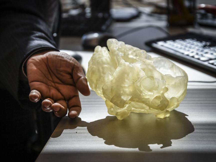
“Without the 3D model, the surgery would be more time-consuming and more stressful. It is very experimental, but a tremendous relief to me. If I can have the model, I’ll know if I can do it, or if it is wise to do it. A better understanding of the 3D spaces will reduce the number of pediatric patients in need of palliative surgery, and in turn, the number of adult patients in need of corrective surgery. This type of surgery is especially applicable to those congenital heart patients age 20-50,” explained Dr. Marcus Haw, the surgical team leader.
Previously, 3D heart models were made using either CT or MRI scans and a program that would turn the data into a 3D model. The heart models were generally useful in surgical pre-planning, although the details available were limited and occasionally imprecise. But interventional cardiologist and congenital heart specialist Dr. Joseph Vettukattil created a new process that combines MRI and 3D echocardiogram data into a single 3D model that has significantly more detail. The MRI data provides a more complete look at the internal structure of the heart and its muscle tissue, while the echocardiogram provides a clearer look at the anatomy of the valves, giving doctors a clearer picture of the heart without needed to examine the real thing.
“A surgeon cannot cut the heart out and look at it. You want to have respect for the heart and cause minimum disturbance and disruption. The surgeon won’t see the back or side because the lungs and other organs are covering it,” Dr. Vettukattil explained.
Borgman holds his own heart in his hands.
The replica of Borgman’s heart was 3D printed in a soft, rubbery material called Tango Plusflx930 on a Stratasys PolyJet 3D printer. The soft material allows doctors to move the walls of the heart around without damaging the model, and more realistically plan their surgical path. Because the doctors were able to create such a clear surgical plan the entire procedure was completed in only two hours–less than half the time it would have taken without the pre-planning. And only six weeks after surgery, the enlarged side of Borgman’s has already started to shrink, and his doctors have cleared him to return to work.
While his new method of generating 3D models of hearts is quite successful, Dr. Vettukattil is still working on more advanced 3D heart models. He wants to incorporate CT scan data in his model so it can generate a more accurate picture of the heart’s external anatomy, especially in relation to the rest of the patients organs. He is also helping to develop a process to use CAD software to create a 3D animated model of the heart in motion. The CAD data could then be viewed through special 3D glasses that allow the doctor to literally see directly inside of the patient’s heart.
Discuss this story in the 3D Printed Heart forum thread on 3DPB.com.
[Source/Photos: MLive.com]Subscribe to Our Email Newsletter
Stay up-to-date on all the latest news from the 3D printing industry and receive information and offers from third party vendors.
You May Also Like
3D Printing Unpeeled: Biofuel Waste to Filament & Sustainable Photopolymers
I can’t ever remember a day with so many potentially high impact news stories have come out. In one story, we all know that there are problems with the safety...
Finnair Hires AM Craft to 3D Print Plastic Parts for Aircraft Interiors
Riga-based AM Craft, a supplier specialized in 3D printing aviation components and certified under EASA Part 21G, announced a significant achievement today. The company will assist in upgrading Finnair’s A320...
3DPOD Episode 198: High Speed Sintering with Neil Hopkinson, VP of AM at Stratasys
Neil Hopkinson, a pioneering 3D printing researcher, played a pivotal role in developing a body of research that is widely utilized today. He also invented High Speed Sintering (HSS), also...
3D Printing Webinar and Event Roundup: May 12, 2024
Webinars and events are picking up in the AM industry this week! ASTM International continues its Professional Certificate Course and Stratasys continues its advanced in-person trainings, while 3D Systems is...