
3D Printed Tadpole Endoscope to Improve Cancer Diagnosis

 There’s a strong human instinct to avoid knowing bad news as if somehow that prevents it from being real. The consequences of succumbing to this kind of irrational desire can range from simply complicating a situation, to finding yourself having lost the opportunity to put up a fight against a fatal disease like cancer.
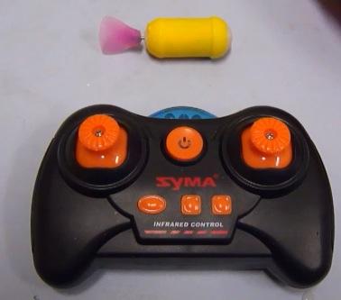
Any number of sources will tell you that when dealing with cancer, early detection is a key element to increasing the chances of survival. In a paper published in the most recent issue of Transactions the journal of the Hong Kong Institution of Engineers (HKIE), a research team from the Chinese University of Hong Kong’s Institute of Precision Engineering described a device they have developed to aid in the early detection of cancers in the gastrointestinal tract.
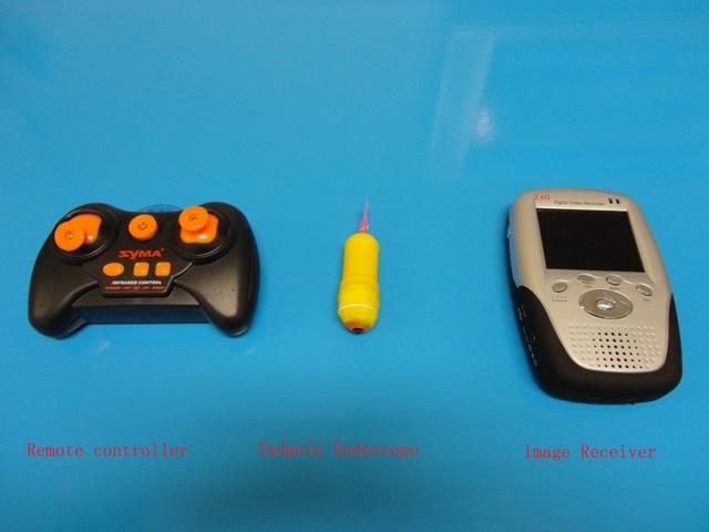
The miniature machine, composed of electronics and a wireless video camera enclosed in a 3D printed shell, is named the Tadpole Endoscope because the mechanics of its movement are based on those used by a tadpole to propel itself through its watery environment. The size of a large pill, the tadpole is introduced into the patient’s gastrointestinal tract by swallowing. Once inside, it can be directed via remote control and send back to a screen images of its surroundings.
 Prior to this, the method for examining the GI tract of a patient was through an endoscopy which consists of the insertion of a flexible tube combined with a light source and an array of lenses or tiny cameras. This tube is then ‘driven’ from the outside and can be unwieldy and difficult to operate in tight areas. In addition, it’s just downright unpleasant.
 Since so much of the battle in cancer lies in convincing people to get themselves examined regularly, removing any aspect of the examination that is uncomfortable, no matter how small, is an important part of the strategy. While it may not seem that the image of a tadpole swimming around in your guts is a vast improvement, anybody who has every experienced an endoscopy probably immediately understands how much better the larval frog approach sounds.
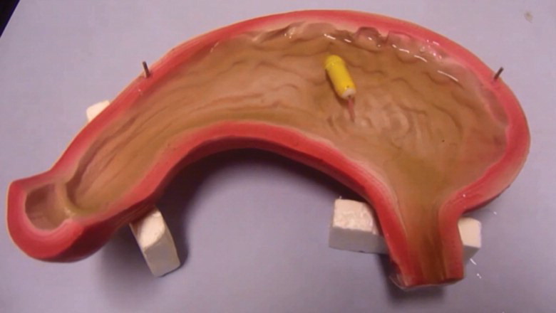
To date, the tadpole has not been released into the GI tract of any human beings. Instead, it has been undergoing rigorous testing, first in a model of the stomach and then later in the stomach of a pig. The successful test of the model with the propulsion system takes it one step closer to being ready for human use, but there are still a great deal more tests to undergo before then. What are your thoughts on this possible breakthrough within the cancer diagnostic space? Discuss in the 3D Printed Tadpole Endoscope forum thread on 3DPB.com.
Subscribe to Our Email Newsletter
Stay up-to-date on all the latest news from the 3D printing industry and receive information and offers from third party vendors.
Print Services
Upload your 3D Models and get them printed quickly and efficiently.


You May Also Like
AMT Shakes Up 3D Printing Market with Affordable, High-Performance Post-Processing Consumables
Additive Manufacturing Technologies (AMT), a global leader in automated 3D printing post-processing, is launching a new line of consumables that promises to significantly reduce operational costs for additive manufacturing users....
The Bambu Lab 3D Printing Platform… or Trapdoor?
Bambu Lab began as a completely closed 3D printing system, where the printer, software, and materials all functioned well but were exclusively from the company itself. This approach mirrored Formlabs,...
2025 Renault 5 E-Tech Electric Is Latest Car with 3D Printed Accessories
Due to the required numbers, additive manufacturing (AM) has struggled to make significant inroads into vehicle interiors in meaningful numbers—at least as far as public knowledge is concerned. Typically an...
BMW Completes Project to Automate Plastic 3D Printing
After a three-year journey to efficiently scale polymer 3D printed part production, the POLYLINE project has concluded. This endeavor, headquartered at BMW’s Additive Manufacturing Campus, pooled the expertise of EOS,...