

Characterization of Conventionally Manufactured & 3D Printed Acetabular Cup Implants


The use of 3D printed porous titanium implants in the orthopaedics field is growing, as they feature a similar design to components manufactured with traditional methods, but have a better bony fixation. A team of researchers from University College London and the Royal National Orthopaedic Hospital published a paper that non-destructively characterizes the dimensional, morphological, and morphometric features of titanium 3D printed acetabular cups.
“3D-printing enables the manufacture of complex porous structures and features that may provide enhanced fixation stability, compared to conventionally manufactured porous geometries [3]. Specifically designed pore shapes can be produced using 3D-printing, unlike traditional technologies where there is a limited control over the porous structure layout [7].The clinical rationale behind the use of 3D-printing for customized (patient-matched) implants was to overcome the limitations of conventional custom components, which could not address complex cases where the bone stock was very limited,” the team wrote.
The goal is to “create porous structures that maximize bone integration,” and 3D printing provides clinicians with more control over the design of the cup, such as making the walls thinner or adding holes with reinforced edges. The cost of the final component is also reduced, since complex assembly and specialized tooling are limited with the use of 3D printing, but there’s still a lot we don’t know about the “potential risks and clinical impact” of 3D printed orthopedic cup implants. In order to gain a better understanding of their mechanical properties, these researchers focused on characterizing the design of the cups.
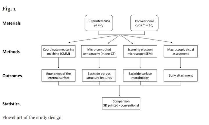
“Using a set of conventionally manufactured cups as reference, we evaluated (1) a dimensional characteristic of the internal cup surface, which could impact the seating of the shell-liner; (2) surface morphology and porous structure on the backside of the cups, which are related to osseointegration; (3) the amount of bony attachment, as first indicator of the bone-implant interaction,” the researchers explained.
They analyzed 16 titanium-aluminium-vanadium (Ti6Al4V) cups, with a median size of 54 mm, that had been retrieved from patients, six of which had been 3D printed using electron beam melting (EBM) technology, with the other ten made by conventional means.
“Patients in the 3D-printed and conventional groups had median (IQR) ages of 61.1 (48.3 to 71.6) and 67.2 (63.3 to 70.1) years (p = 0.5941), a median (IQR) time to revision of 24.9 (21.1 to 46.8) and 49 (23.8 to 62.1) months (p = 0.296) and genders were 20% and 43% male, respectively,” they wrote.
The researchers used a photogrammetric method to determine the amount of bony attachment on the cups, which was only marked when they could see a white porous structure. Coordinate-measuring machine (CMM), electron microscopy (SEM) and microcomputed tomography (micro-CT) were used to investigate the following:
- roundness of internal cup surface
- morphology of backside surface
- morphometric features of the porous cup structures
The micro-CT analysis was a three-step process, starting with scanning the acetabular components and reconstructing the data, segmentation and volumetric rendering of the data with software, and measuring the porous structures’ morphometric parameters.
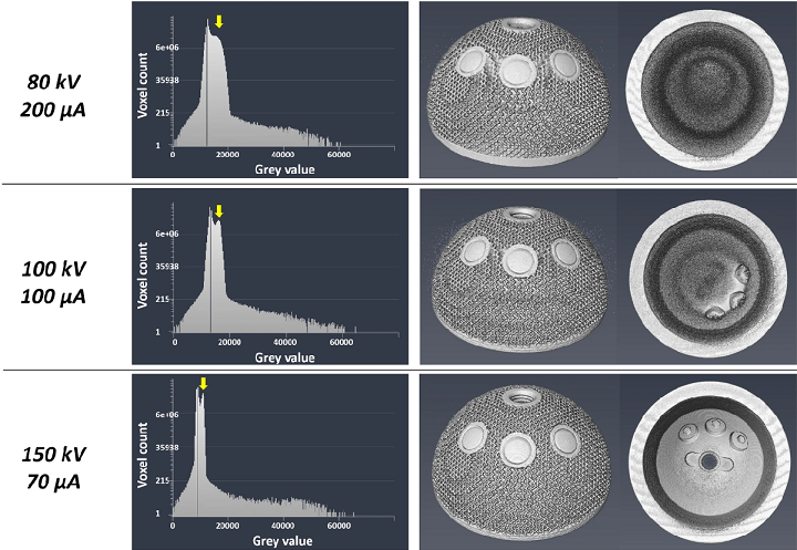
Optimization process outcomes of micro-CT scanning parameters. From top to bottom, the investigated parameters were 80 kV, 200 μA, 100 kV, 100 μA and 150 kV, 70 μA. 150 kV, 70 μA was chosen because the peak corresponding to the metal (yellow arrow) in the voxel count histogram was higher compared to the same peak in the plots related to other parameters. This suggested a better separation between voxels belonging to the background (air) and voxels belonging to the material.
The researchers used the 3D reconstructed models of the cups to “assess clinically relevant morphometric features” of the porous structure. These features were porosity, pore size, and strut thickness, and the first two “represent the available space for bone ingrowth,” while the site for bone cell attachment is the strut thickness. They also measured the porous structures’ thickness, which represents “the maximum penetration depth for bone tissue.”
“Statistical analysis was performed using Prism 7 (GraphPad, USA). The differences between 3D-printed and conventional cups in terms of roundness of the internal cup surface, morphometric features of the porous structures (porosity, pore size, strut thickness), thickness of the porous structure and bony tissue attachment were evaluated using non-parametric Mann-Whitney tests,” the researchers wrote.
The results of the CMM showed a median roundness of 19.45 and 14.52 μm for both the 3D printed and conventional cups, and “the SEM analysis revealed differences in surface morphology” between the backside surfaces of the two types of cups, which the researchers had expected.
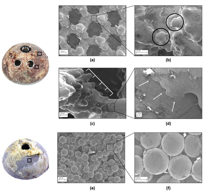
SEM images of 3D printed and conventional implants. a) 3D printed porous structure had a regular shape with clearly identifiable pores; b) partially molten particles (black circles) were visible on the struts; c) the ‘stair-step’ effect due to 3D printing; and d) texture lines also found at higher magnification. Conventional cups showed e) less clearly identifiable pores and f) beads of uniform distribution along the backside surface.
They saw partially molten particles, with a median size of 73 μm, and the stairstep effect, on the 3D printed implants’ struts, both of which are by-products of the method. But, the conventional cups had less visible pores between the packed beads, which is not one.
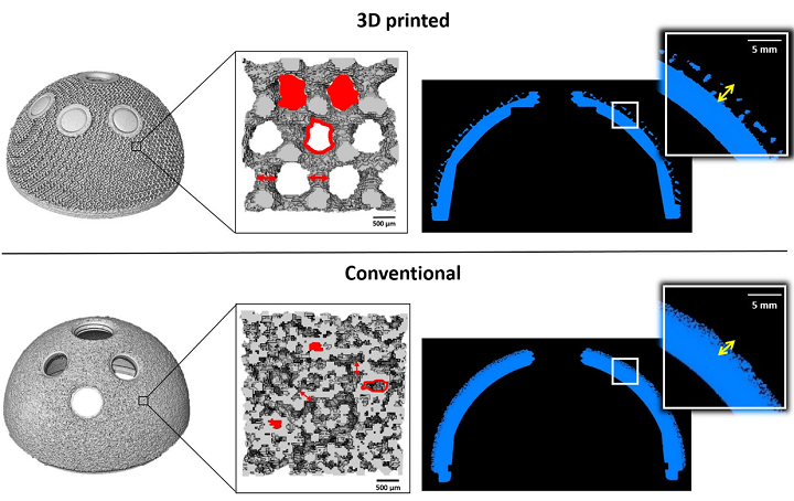
Outcomes from micro-CT analysis of 3D printed and conventional cups. L-R, the reconstructed implants and a zoom on the porous structures with the morphometric parameters depicted are exhibited. Porosity is defined as the percentage of void volume (red filled shapes) over total volume; pore size is the diameter of the circle whose area equals one of the red empty shapes; and strut thickness is indicated by red arrows. A cross-section of the components and a zoom showing the thickness of the porous structure (yellow arrows) are also shown.
“As expected, porosity, pore size, strut thickness and thickness of the porous structure were significantly higher for 3D-printed components (p = 0.0002), with median values of 72.3%, 915 μm, 498 μm and 1.287 mm (p = 0.0002),” they wrote.
You can see the median measurements of the morphometric features for the “backside porous structures” of the cups in the table below.
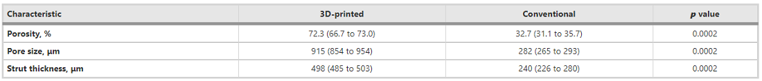
Median measurements of the morphometric features of the porous structures of both types of implants.
The porosity of the 3D printed cups was twice that of the other kind, while the strut thickness making up the porous framework of the conventional cups “was half the dimension exhibited by 3D-printed implants.” The pores of the conventional cups had a diameter three times smaller than the 3D printed ones, which had a thicker porous structure.
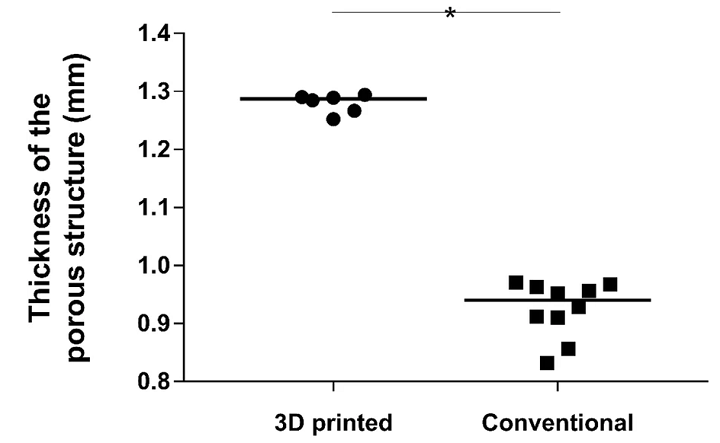
Dot plot showing distribution of values of thickness of the porous structure measured for the groups. The solid line represents the median value.

CT cross-section slice of a 3D printed cup with grey areas (white arrow) depicting presence of a material different from the Ti6Al4V alloy and background air.
“The 3D-printed and conventional cups had a median (IQR) bony attachment surface coverage of 84.9% (65 to 95.6) and 69.3% (65.1 to 93.9), respectively (p = 0.2635),” the researchers wrote about the results of the macroscopic visual assessment.
Due to a “comparable roundness of the internal surface” of all the cups, they determined that the dimensional property was not influenced by the two types of manufacturing, and both groups of cups “exhibited the presence of particles.”
“In general, these morphological features provide a rough surface which may promote osteoblast cell adhesion and growth (osteoconduction),” they stated.
“This falls under the material ‘contact guidance’ principle, whereby the cell movement and development is affected by the surface morphology of the implant [37].”
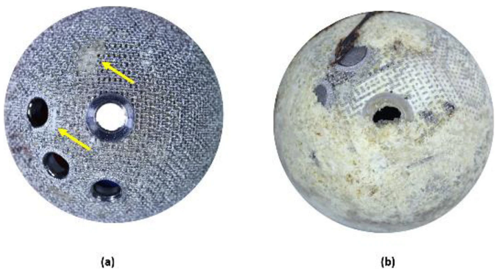
Example of low (a) and high (b) percentage of bony attachment on 3D printed cups. The presence of bone is indicated by the yellow arrows.
The researchers explained that the basic goal of any cementless acetabular cup design is good primary stability and long-term bone fixation, and that the 3D printed cups they studied “showed morphometric values similar to those of trabecular bone.”
“Interestingly, we identified a third material, different from Ti6Al4V and background air, in the cross-section slices of the 3D-printed cups obtained from the micro-CT investigation. These preliminary findings suggest that this may be the bone that grew into the porous structure of the cups, because only a material dense enough to attenuate the incoming X-rays would be visible in the CT slices.”
It should be mentioned that the study was limited due to the fact that the team only considered 3D printed cups made with EBM technology, and none that were fabricated with selective laser melting (SLM) 3D printing, which has also been used to make acetabular cups.
“Future work involving larger and more heterogeneous cohorts of implants are needed in order to understand if the transition to 3D-printing techniques for mass production of orthopaedic implants can provide safe and well-performing outcomes,” the researchers concluded.
Discuss this and other 3D printing topics at 3DPrintBoard.com or share your thoughts below.
Subscribe to Our Email Newsletter
Stay up-to-date on all the latest news from the 3D printing industry and receive information and offers from third party vendors.
Print Services
Upload your 3D Models and get them printed quickly and efficiently.


You May Also Like
3D Printed Chip Packaging Specialist XTPL Enters Japanese Market
The additive manufacturing (AM) industry naturally wants to move beyond prototyping to production at scale, and the industry is certainly starting to demonstrate success with that objective, especially in Asia....
UT Researchers Use 3D Printing to Develop “Tabletop EUV Lithography” Process
Photolithography, the semiconductor manufacturing process whereby lasers transfer patterns onto chemical layers coating a substrate, is one of the most amazing industrial processes humanity has ever created. It is also...
3D Printing News Briefs, May 30, 2026: RIMPAC 2026, Acquisition, Ceramic Implants, & More
We’re kicking things off with materials news in this weekend’s 3D Printing News Briefs. Then it’s on to a hybrid manufacturing system for a maritime exercise, an expansion of industrial...
The University of Utrecht: “3D Printing Could Change Who Gets to Become a Manufacturing Power”
For decades, manufacturing has mostly been controlled by countries with huge factories, lower labor costs, and industrial systems that took years, sometimes decades, to build. But Utrecht University human geographers...


