

3D Printed Monolithic Device Acts as Trap to Isolate Cancer Cells


International researchers have recently developed a method for capturing blood cells to isolate tumors, using a novel liquid biopsy device that functions as a 3D printed cell trap.
This technique is being used in research for cancer treatment as an improved way to target cancer cells, which can be difficult to find (and often to allow for a diagnosis)—as well as monitoring for the further spreading of cancer cells if they metastasize.
To put it into perspective, it is important to understand that the few cancer cells being sought could be hidden in billions of other blood cells. The 3D printed trap finds these cells as white blood cells are kept in, and red blood cells are filtered back out. While previous methods have been used, in most cases they lack efficiency and damage cells in the trap.
“Capturing these tumor cells is in itself a big challenge,” says researcher Fatih Sarioglu. “Because there are billions of blood cells, you need an engineering tool, a technology that can screen the cells one by one. You cannot miss even one cell. Typical lab techniques don’t work.
“The other problem is that cancer cells constantly mutate, so you can’t rely on one type of marker to distinguish them from the other cells, even from blood cells, and they can sometimes hide very efficiently between the blood cells.”
Made up of 32 microfluidic layers, the cell trap measures 100 mm x 20.5 mm x 19.2 mm and is fully detailed in the recently published ‘Hybrid negative enrichment of circulating tumor cells from whole blood in a 3D-printed monolithic device’, featuring Chia-Heng Chu as the lead author.
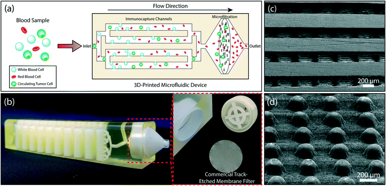
The design of the 3D printed microfluidic device. (a) A schematic showing the tumor cell enrichment process in the device. Whole blood is introduced to the device. WBCs are captured in the multi-layered immunocapture channels. A membrane filter retains all nucleated cells (including the residual WBCs) and eliminates anucleated blood cells. (b) A photo of the 3D printed device showing the microfluidic channels with 32 stacked microfluidic layers and the filter holder (right). The membrane filter can be accessed by removing the threaded cap (left). (c) A scanning electron micrograph of the cross-section of the device showing 200 μm-diameter microposts within the microfluidic layers. (d) Arrayed micropillars, within each layer, are shifted by 10 μm from row-to-row to maximize cell-micropost interactions.
The device functions with two different sections: a multi-layered immunoaffinity-based leukocyte capture section and a filtration section, with the design created in SolidWorks and then 3D printed on a ProJet 3510 HD 3D printer with VisiJet® M3-X plastic material. Device size was restricted due to the size of the centrifugation tube required for dewaxing.
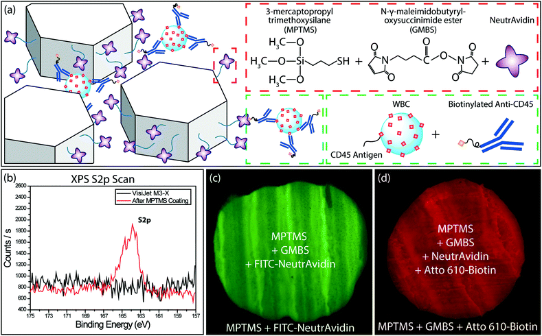
Surface functionalization of the 3D printed material and characterization. (a) A schematic showing the (top right) surface functionalization and (lower right) WBC labeling chemistries. (b) Results from the XPS scan of sulfur on the surface of VisiJet® M3-X material before and after the coating of MPTMS. The peak confirms the successful coating. Fluorescence microscope images of (c) NeutrAvidin (FITC) and (d) biotin (Atto 610) on chemically modified VisiJet® M3-X material. Differential fluorescence between the functionalized surface and the exterior control surface confirms the specific surface modification.
3D printing allowed for the more streamlined fabrication of the device, especially due to the potential for a larger surface area; however, the technology also presented a major issue in the need to eliminate the solid wax used as a necessary ‘sacrificial filler.’
“We tried a bunch of different ways using traditional medical tools in bio labs and ended up with a standard centrifuge that removes the hard wax by heating the trap and spinning it to extract the then liquid wax from the channels,” said Sarioglu. “That was a manufacturing challenge that we had to work on.”
Simulated samples were created as they ‘spiked’ tumor cells into whole blood, mainly culturing Ovarian cancer cell line HeyA8, human breast cancer cell line MDA-MB-231 (ATCC® HTB-26™) and prostate cancer cell line LNCaP (ATCC® CRL-1740™).
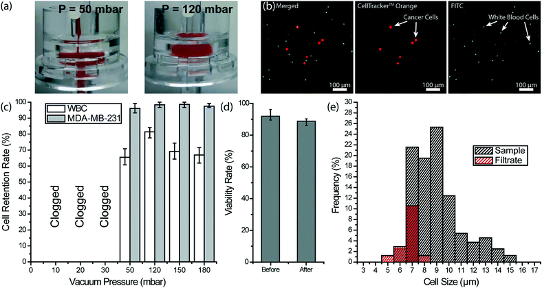
Removal of RBCs and platelets from whole blood with a membrane filter. (a) Images of a 3 μm-pore membrane filter operated under different buffer pressures to recover spiked cancer cells from whole blood. (left) When the filter was operated at 50 mbar, a cake layer was formed due to RBC accumulation. (right) At 120 mbar, RBCs squeezed through pores more efficiently and no cake layer could be observed. (b) Fluorescence microscope images of nucleated cells retained on the filter. The MDA-MB-231 breast cancer cells were pre-stained with CellTracker™ Orange and the WBCs were labeled with FITC-conjugated anti-CD45 antibody. (c) Measured retention rates for both spiked MDA-MB-231 breast cancer cells and the WBCs under different vacuum pressures. (d) Measured MDA-MB-231 tumor cell viability rate before and after processing through the membrane filter under 120 mbar. (e) Measured cell size distribution of the WBCs in the sample and the filtrate at 120 mbar vacuum pressure showing that no WBCs larger than 8 μm were able to pass through the membrane filter.
Post-filtration of the ‘leuko-depleted blood’ made it possible for the researchers to maintain all the nucleated cells—even leftover WBCs that were on the detachable membrane filter. Overall, the team sees the potential for further advancement and even better performance in their scalable technique.
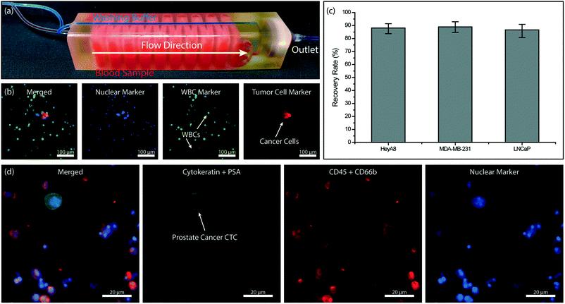
Tumor cell enrichment from whole blood using the full device. (a) A photo of the full 3D printed device. The device is filled with red- and blue-colored dyes to visualize dedicated sample and buffer paths within the device, respectively. (b) Fluorescence microscope images of the nucleated cells retained on the membrane filter. Hoechst 33342 dye was used to stain the cell nucleus. (c) Measured recovery rates for ovarian (HeyA8), breast (MDA-MB-231) and prostate (LNCaP) cancer cells spiked into whole blood samples. (d) Fluorescence microscope images of the prostate cancer CTC retained on the membrane filter.
“This is what I am living for in a way: You spend time to think of something, work hard to realize it, and at the end you see it working with the idea that it will help people,” said Sarioglu. “That’s why I made the transition from just building sensitive tools and engineering devices to things that can help people and impact their lives.”
A variety of 3D printing devices are being used these days for diagnostics and treatment of cancer, along with training devices like 3D printed phantoms. What do you think of this news? Let us know your thoughts! Join the discussion of this and other 3D printing topics at 3DPrintBoard.com.
[Source / Images: ‘Capturing Blood Cells to Isolate Tumor Cells’; Hybrid negative enrichment of circulating tumor cells from whole blood in a 3D-printed monolithic device’]Subscribe to Our Email Newsletter
Stay up-to-date on all the latest news from the 3D printing industry and receive information and offers from third party vendors.
Print Services
Upload your 3D Models and get them printed quickly and efficiently.


You May Also Like
Divergent Declares that German 3D Printers are Superior, And Plans Massive LPBF Expansion
Divergent has announced a new version of its Laser Powder Bed Fusion (LPBF) printer and a new site. The company aims to do nothing short of “further accelerating its mission...
Incodema3D Buys 14 Metal EOS Systems, Now One of the World’s Largest Metal 3D Printer Operators
Recently, a majority stake of 3D printing service bureau Incodema3D was purchased by AFM Capital. Under new ownership, the Freeville, New York company is now using its cash-rich parent for...
CEO Yoav Zeif on Why Stratasys’ Markforged Acquisition Is Really a Bet on Industrialization
When Stratasys announced plans to acquire Markforged, the immediate focus was on the deal. Markforged is one of the most recognizable names in additive manufacturing (AM), known for its continuous...
3D Printing & the Autonomous Era: Defense Tech’s Latest Mutation
When we last checked in on the broad defense tech landscape and the role of the additive manufacturing (AM) industry in that environment, it became clear that the connecting thread...


