
UCLA & Swedish Researchers Create 3D Printed Microscope Attachment to Aid in Field Diagnostics for Cancer


 Once again, 3D printing is allowing for impacts in the medical field that most of us could not have begun to comprehend just several years ago. While we report on many new medical devices helping patients around the world, the latest is a smartphone device created by researchers at California NanoSystems Institute at UCLA and Swedish scientists from both Stockholm University and Uppsala University.
Centering around DNA research, the teams have found a way to make it easier to run DNA sequences and analysis. Again, 3D printing is part of what makes this possible due to affordability in creating a new and durable device that can be used in the field—especially important in developing countries and communities lacking in resources. With a microscope that is smartphone based and completely portable, health care workers will be able to get fast results without the lab—or the waiting.
This new device is discussed in ‘Targeted DNA sequencing and in situ mutation analysis using mobile phone microscopy,’ by Malte Kühnemund, Qingshan Wei, Evangelia Darai, Yingjie Wang, Iván Hernández-Neuta, Zhao Yang, Derek Tseng, Annika Ahlford, Lucy Mathot, Tobias Sjöblom, Aydogan Ozcan, and Mats Nilsson.
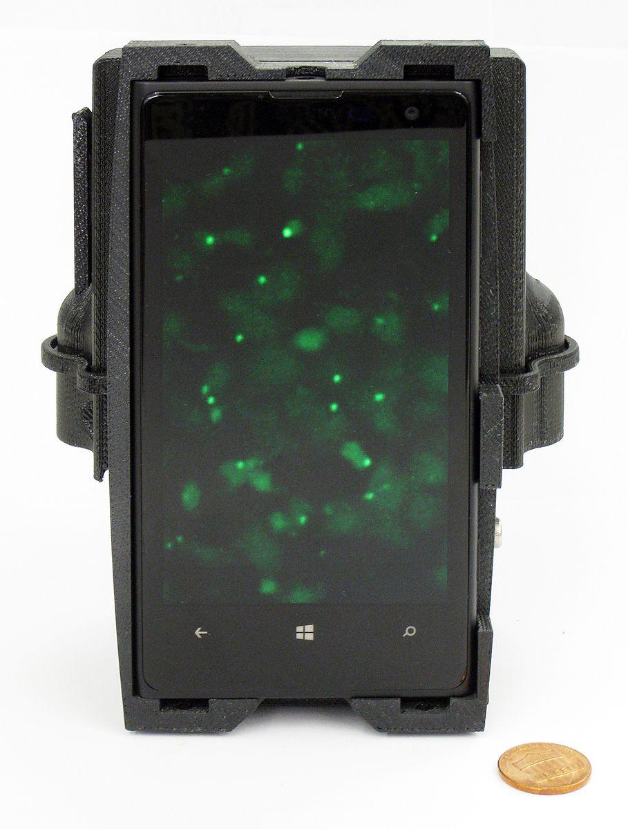
[Photo credit: UCLA, Stockholm University and Uppsala University]
“… we designed and 3D printed a light-weight optomechanical attachment that is integrated with the existing camera module of a mobile phone. This optical attachment contains two compact laser diodes (at 532 and 638 nm) for multicolour fluorescence imaging and a white light-emitting diode (LED) for bright-field transmission imaging.”
Aydogan Ozcan, UCLA’s Chancellor’s Professor of Electrical Engineering and Bioengineering and an associate director of CNSI, is one of the team leaders on the research. He states that if they can get these devices into mass production, they should cost less than $500 each.
“A typical microscope with multiple imaging modes would cost around $10,000, whereas higher-end versions, such as the one we used to validate our mobile-phone microscope, would go for $50,000 or more,” Ozcan said.
Diagnostics obviously play a large part in detecting disease, and as that process becomes further refined patients will continue to benefit.
“It’s very important to have these molecular testing approaches at a doctor’s office or where care is being given,” said Mats Nilsson, also a leader on the project, and a professor of biochemistry and biophysics at Stockholm and Uppsala Universities and the director of SciLifeLab in Stockholm. “Oftentimes, advanced lab-based testing is performed at major hospitals, which is limiting, as not everyone has access to a hospital that can perform these tests.”
Technicians are able to analyze DNA easily while out in the field through a simple process. After taking a tissue sample and placing it in a container, they allow the microscope to take images which are then analyzed via data being fed to an algorithm. With this process, they are able to spot mutations inside a tumor, for example. According to the researchers, even small groups of cancer cells can be detected.
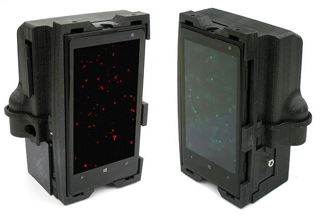
[Photo credit: UCLA, Stockholm University and Uppsala University]
“Ultra–low-cost DNA sequencing and tumor biopsy analysis, in which morphology and mutation analysis are combined, can substantially decrease diagnostic costs and make it more widely accessible,” said Malte Kühnemund, one of the lead authors on the paper.
According to UCLA Newsroom, this research was supported by the National Science Foundation, the Office of Naval Research, the Army Research Office, the Howard Hughes Medical Institute, the Swedish Research Council, the Swedish Cancer Foundation, SciLifeLab and OncoTrack, a project of Europe’s Innovative Medicines Initiative. Discuss in the 3D Printed Microscope Attachment forum at 3DPB.com.
[Source: UCLA]Subscribe to Our Email Newsletter
Stay up-to-date on all the latest news from the 3D printing industry and receive information and offers from third party vendors.
Print Services
Upload your 3D Models and get them printed quickly and efficiently.


You May Also Like
Excellent Desktop Injection Molding, Made in Italy by Robot Factory
I was captivated when I saw my first Robot Factory 3D printer. The robust, precise machine was built to last. And this was in an era of very flimsy, disposable,...
Pogačar & Fairlight Cycles Show Us Low Cost 3D Printed Components for Bikes
There has been a lot going on in 3D printing for bicycles over the years. The most successful implementation so far is in bicycle seats. Carbon 3D printed seats are...
3D Printing News Briefs, June 18, 2026: Reseller, Relocation, Metal Space Powder, & More
We’ll start with business news in today’s 3D Printing News Briefs, as XJet appointed a value-added reseller in Germany, BIO INX is expanding its presence in the Italian market, and...
Researchers Combine AI and Bioprinting to Create Tiny Blood Vessel Networks
If 2026 has a theme in bioprinting, it may be blood vessels. Researchers can already print incredibly sophisticated tissues. The harder part is keeping those tissues alive. Without a network...


