
Researchers 3D Print Breast Phantoms for Better Training in Cancer Diagnosis


US researchers are looking for ways to improve the diagnosis of breast cancer and the use of phantoms, outlining their findings in the recently published ‘Imaging properties of 3D printed breast phantoms for lesion localization and core needle biopsy training.’
A core needle biopsy (CNB) often accompanies the process of diagnosing breast cancer, due to excellent accuracy; however, like any other technology, training is required for radiologists. The use of phantoms in training continues to be successful, allowing for hands-on education regarding procedures that involve imaging probes and procedural instruments. Affordability is a limiting factor though, with commercial biopsy phantoms typically priced from $350 to $450. Because of this, many training programs use alternative materials like food, imitating soft tissue.
In the development of 3D printed breast phantoms for this study, the researchers realized the ideal product would offer the following features:
- Anatomical accuracy
- Adequate visualization of target lesions
- Tissue integrity
- Potential for several uses
- Easy maintenance
- Affordability
Previous research studies have yielded a variety of 3D printed phantoms for purposes in bioprinting, dosimetric verification, cardiac health, and more.
“In the context of breast imaging, several papers have explored the benefit of 3D printed phantoms for image quality analysis, surgical planning, and implantable bioprinted breast scaffolds,” stated the authors. “Researchers have also created phantoms from 3D printed molds filled with ultrasound compatible polymers. However, to our knowledge, no 3D printed phantoms which accurately mimic breast tissue characteristics, are feasible for multimodality imaging, and are produced at a low cost, exist.”
Three samples were created, customized for both training in performing biopsies as well as imaging evaluation. Traditional phantoms were also fabricated, using chicken breasts, gelatin, and olives and blueberries to mimic tumors.
Breast Phantom quality assessment
Each phantom was assessed by a trained breast radiologist. They were also tested in biopsy training seminars (consisting of at least four trainees each time), rated on a scale of one to five.
Evaluations showed that the 3D printed breast phantoms were more accurate than the traditional models, with the TissueMatrix material demonstrating the best tactile simulation of tissue. The researchers did note, however, that there was a ‘pronounced limitation’ due to the lack of US beam penetration.
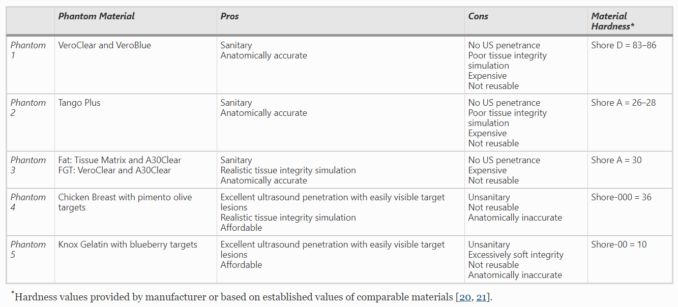
3D printed Breast Phantoms. Phantoms were printed in VeroClear (a), TangoPlus (b) and a new combination of TissueMatrix with a coating of VeroClear at 600 μm (c). Ultrasound (US) image (d) of the TangoPlus phantom demonstrates lack of sound penetration. Images obtained with standoff pad (e) shows similar findings. Both VeroClear and TissueMatrix phantoms had similar results on US imaging
The chicken breast phantom showed the best results in terms of US beam penetration, image quality, and material hardness. The phantom made from gelatin displayed ‘acceptable beam penetration and image quality,’ but inferior tactile stimulation quality.
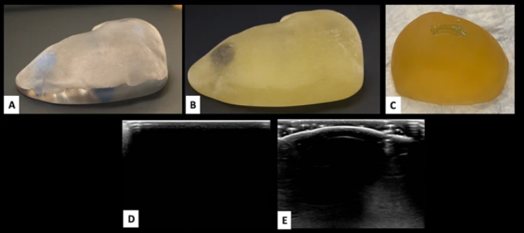
Fragility of Knox gelatin phantom. Knox gelatin breast phantom (a) easily fragments with excessive pressure. Ultrasound images after several biopsy attempts create linear air tracts (b and c), reducing visibility of target lesions
“Gelatin is particularly fragile and the density too soft compared to human breast tissue. Small cracks created in the gelatin during routine handling, multiple biopsies, or excessive pressure from the probe can create pockets of air within the model and obstruct visualization of targets,” added the researchers in conclusion. “The short shelf life of the gelatin and creation of air tracks with each biopsy limits reusability. Preservatives may be added to extend shelf life; however, the single use nature of gelatin models makes preservatives unnecessary.
“Limitations of this study include the lack of additional types of printing technologies or materials, including flexible materials printed with the use of material extrusion, vat photopolymerization, and other types of material jetting printers (i.e. 3D Systems and Mimaki).”
What do you think of this news? Let us know your thoughts! Join the discussion of this and other 3D printing topics at 3DPrintBoard.com.
[Source / Images: ‘Imaging properties of 3D printed breast phantoms for lesion localization and core needle biopsy training’]Subscribe to Our Email Newsletter
Stay up-to-date on all the latest news from the 3D printing industry and receive information and offers from third party vendors.
Print Services
Upload your 3D Models and get them printed quickly and efficiently.


You May Also Like
3D Printing News Briefs, July 9, 2026: RIMPAC 2026, Software, Housing, & More
In today’s 3D Printing News Briefs, Massivit continues its focus on aerospace and defense manufacturing, and Meltio is collaborating with Phillips Corporation for RIMPAC 2026. Moving on to software, AMIS...
3DPOD 304: Precast Concrete AM with Greg Kerkstra, Mangrove
Greg Kerkstra is part of a family business that leads in the precast concrete industry. They’ve now turned to Progress Group’s large-format binder-jet concrete technology, which we covered here in...
3D Printing News Briefs, June 10, 2026: Grand Opening, Photoresins, Footwear, & More
We’re starting with some exciting news in today’s 3D Printing News Briefs: Stratasys just celebrated the opening of its new North American headquarters in Minnesota. Moving on, Nanoscribe is scaling...
Wells Fargo Backs ICON in Landmark Milestone for 3D Printed Housing
Qualification is an indispensable step on the path to legitimization for any new technology, but it’s still just one step: markets tend to remain unswayed without a co-sign from an...


