Could Transparent Human Organs Help Create Artificial 3D Printed Ones?

Imagine being able to understand and see the complex structures of human organs at the cellular level. On February 13, a group of researchers in Germany’s Ertürk Lab, in the Institute for Stroke and Dementia Research (ISD) at Ludwig Maximilian University Hospital in Munich, published a paper based on a new technology that can make intact human organs transparent. Thanks to microscopic imaging they could visualize underlying complex structures of see-through organs at the cellular level, resulting in organ maps that could serve as templates for 3D bioprinting technologies, as well as eventually contribute to the creation of on-demand artificial organs for patients in need. Furthermore, the researchers suggest that tissue engineering efforts demand cellular maps of human organs to replicate large-scale human tissues and organs by emerging technologies including 3D bioprinting.
It was all possible thanks to the development of a new technology called Small-micelle-mediated Human orgAN Efficient clearing and Labeling (SHANEL) which allowed researchers to chart the cellular and molecular architecture of large intact mammalian organs. Genetics and neuroscience specialist Ali Ertürk, director of the Institute for Tissue Engineering and Regenerative Medicine (ITERM) at the German Research Center for Environmental Health and also principal investigator at the ISD, published the findings of the groundbreaking project that used microscopic imaging technology to reveal underlying complex structures of human organs by making them see-through in the scientific journal Cell.

Vascular and glomeruli details of the human kidney
Along with 22 research colleagues, including Shan Zhao, Ingo Bechmann, Marco Duering, Oliver Bruns, Bjoern Menze, Victor Puelles, Jan Lipfert, and Eckhard Wolf, they created SHANEL, a robust and unbiased technology based on a new tissue permeabilization approach to clear and label stiff human organs and chart the cellular and molecular architecture of large intact mammalian organs. The team of scientists used the new approach to deliver a transparent intact adult human brain and kidney and perform 3D histology with antibodies and dyes in centimeters-depth. Thereby, revealing structural details of the intact human eye, human thyroid, human kidney, and transgenic pig pancreas at the cellular resolution.
Ertürk suggested in his Twitter account that “mapping the human brain or other organs is a crucial step forward deciphering how they function in health and disease.” He also asserted his belief that “SHANEL can help to map the human brain at the molecular level and provide cellular blueprints of human organs for 3D bioprinting technologies to make new organs on demand.”
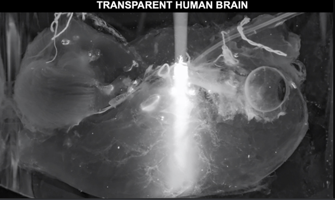
Light of an epifluorescent microscope can travel in the brain end-to-end, demonstrating the full transparency of intact human brain
According to news released by the German Research Center for Environmental Health, deciphering the structural complexity of human organs has always been a major challenge due to the lack of technologies to image them at the cellular level. The publication suggests that the progress in mapping intact human organs has been limited, especially in deciphering the anatomical complexity, mainly due to a lack of scalable technologies to image human organs at the cellular level. Although magnetic resonance imaging (MRI) can provide longitudinal imaging for human organs including the brain and kidney, it lacks cellular resolution.
The researchers had recently made developments in tissue clearing that allowed them to obtain first cellular views of intact transparent mouse organs in 3D. In the previous work, the researchers applied a technique that made dead mice transparent and hard like plastic, giving researchers an unprecedented view of how different types of cells interact in the body. The approach allowed scientists to pinpoint specific tissues within an animal while scanning the entire body. However, working with human organs was not the same, since they are particularly stiff due to the accumulation of insoluble molecules including collagen in tissues that have grown for years or even decades.
To overcome the challenge, they couldn’t use the traditional detergents that were being used for making mouse organs transparent because they didn’t work on human organs, particularly adult ones. They reveal that after exhausting trials, the team discovered that a detergent called CHAPS forms smaller micelles allowing full permeabilization of aged human organs and could make small holes throughout the entire stiff organs. CHAPS allows additional solutions to travel deep into centimeters-thick human organs and convert them into a transparent structure.
“We had to change our approach completely and start from scratch to find new chemicals which can make human organs transparent,” said Shan Zhao, a doctoral student at the German Research Center for Environmental Health and first author of the study.
“Tissue Clearing methods work well on rodent tissue but haven been poorer on stiff and aged human organs. Making the whole human organs transparent required a new approach. We identified CHAPS, a detergent that can permeabilize the aged human organs. Further, we used Acetic Acid and Guanidine Hydrochloride to enhance the depth of molecular labeling,” continued Ertürk in his Twitter account.
The expert also suggested that working with LaVision BioTec, a German-based developer and manufacturer of advanced microscopy solutions for research in neuroscience, immunology, oncology, and developmental biology, helped them create a prototype light-sheet microscope with an extended stage to image human organs as large as the kidney (with a size of 11.5 cm x 8.2 cm x 3 cm). The team also developed deep learning algorithms to analyze hundreds of millions of cells from brain scans in a fast and accurate way.
Moreover, after making the human organs transparent, which were obtained post mortem from Bechmann’s lab at the University of Leipzig, in Germany, the team had to tackle additional challenges for both organ imaging and the analysis of a large amount of resulting data. First, they developed a new laser-scanning microscope with a large sample holding capacity called “Ultramicroscope Blaze” in collaboration with Miltenyi Biotec. This microscope enabled the imaging of human organs as large as the kidney. Next, together with Bjoern Menze from the Technical University of Munich (TUM), the team developed deep learning algorithms to be able to analyze hundreds of millions of cells in 3D.
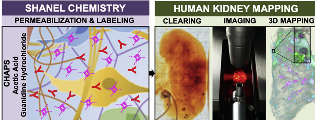
The SHANEL Method
Ertürk also described some shortcomings which he hopes they will correct in the future: the method cannot eliminate the high autofluorescence of human tissue; there are still no light-sheet microscope systems to scan the human brain; SHANEL pretreatments work only with partial commercialized antibodies for deep tissue labeling; the labeling and clearing process still take months depending on the size of the organs; their machine learning algorithm is designed only for the analysis of cell bodies while new algorithms are needed for each structure, such as vessels and neurons, and the best human organ mapping would require very fresh organs, like organs shortly after death, which are very difficult to come by.
The work was conducted at the ISD, the German Center for Environmental Health, Ludwig-Maximilians University Munich, TUM, and supported by The German Research Foundation, NVIDIA, Fritz Thyssen Foundation, National Institutes of Health and The European Research Council (ERC).
In the past few years, scientists have attempted to create methods that can make an entire organ clear allowing the study of the internal structures. This new technique produces incredibly detailed 3D maps of organs, thanks to special dyes added to them in a now-transparent state, allowing researchers to map kidneys, eyes, and brains on a cellular level, which could one day help scientists 3D print the organs. For example, they suggest that understanding the 3D structure of the human kidney would be very valuable for tissue engineering efforts aiming to generate artificial kidneys using 3D bioprinting technologies that require detailed cellular and molecular knowledge on the intact human kidney to be replicated.
Now, thanks to SHANEL, looking at the cellular 3D maps of a condensed light traveling end-to-end in a transparent human organ has such an intricate level of detail which makes it otherwordly and in the future could become a stepping stone for scientists trying to biofabricate organs.
“There is a huge shortage of organ donors for hundreds of thousands of people,” indicated Ertürk. “The waiting time for patients and the transplantation costs are a real burden. Detailed knowledge about the cellular structure of human organs brings us an important step closer to creating functional organs artificially on demand.”
(Images: Helmholtz Zentrum München, Ertürk lab)
Subscribe to Our Email Newsletter
Stay up-to-date on all the latest news from the 3D printing industry and receive information and offers from third party vendors.
You May Also Like
Gorilla Sports GE’s First 3D Printed Titanium Cast
How do you help a gorilla with a broken arm? Sounds like the start of a bad joke a zookeeper might tell, but it’s an actual dilemma recently faced by...
Nylon 3D Printed Parts Made More Functional with Coatings & Colors
Parts 3D printed from polyamide (PA, Nylon) 12 using powder bed fusion (PBF) are a mainstay in the additive manufacturing (AM) industry. While post-finishing processes have improved the porosity of...
$25M to Back Sintavia’s Largest Expansion of Metal 3D Printing Capacity Since 2019
Sintavia, the digital manufacturing company specializing in mission-critical parts for strategic sectors, announced a $25 million investment to increase its production capacity, the largest expansion to its operations since 2019....
Velo3D Initiates Public Offering in a Bid to Strengthen Financial Foundations and Drive Future Growth
Velo3D (NYSE: VLD) has been among a number of publicly traded 3D printing firms that have attempted to weather the current macroeconomic climate. After posting a challenging financial report for 2023,...