
CASMER: Medical Imaging Mannequin Features 3D Printed Organ Models


Canadian researchers used 3D printing to create anatomically correct models of human organs, detailing their findings in the recently published ‘3D Printed CT-based abdominal structure mannequin for enabling research.’ Attempting to improve the medical imaging process, as well as offering more comprehensive education, the team created 3D printed organ ‘shells’ for the mannequin.
When treating patients with radiation, it is critical that errors are not made, exposing them to added, harmful radiation. The authors point out how important it is, in fact, to minimize radiation while maximizing diagnostic data from scans. Anthropomorphic X-ray phantoms are created in the form of the human body to detail areas to be targeted by radiation but can also be critical tools for training of medical professionals.
Phantoms created via traditional methods today are not only limited, but they are cost-prohibitive to produce.
“There has been a particular lack of modular anthropomorphic abdominal phantoms that allow the user to remove and replace the organs to replicate different pathologies, and if required, to place foreign bodies such as dosimeters or surgical devices inside the abdominal cavity,” explain the researchers. “Advances in 3D printing technology have increased the range of possibilities in the creation of innovative models for medical purposes.”
With 3D printing comes the benefit of being able to fabricate organ models that are accurate, and removable. When deciding on materials, however, users must be discerning in terms of structural properties, mechanical properties, and radiological properties defining interaction with the X-rays.
The sample CT-based abdominal structure mannequin, also known as CASMER, featured both 3D printing of almost all the organ shells, along with added packing material to flesh out the anatomy correctly. The 3D printing was no casual feat either, requiring numerous steps, and input from the following:
- Radiologists
- Technologists
- Physicists
- Biomedical engineers
Four different techniques were used:
- Realistic 3D printing of abdominal organs
- Material-based molding of the pancreas
- Beeswax sculpting of abdominal fat
- ‘Off-the-shelf parts for the skeleton and outer shell
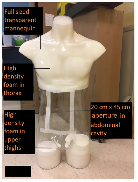
Muscle was made from Clear Flex® urethane rubber (Smooth-ON, PA), while fat was created from modeling beeswax. The mannequin, a ‘hollow polycarbonate full-body’ model, held the 3D printed organs, along with sample bones and muscles, and a pancreas too.
3D printing of the abdominal organs
“The polycarbonate shell was confirmed to minimally attenuate the X-ray radiation from the CT scan, and was transparent to visible light, which facilitated visualization of the internal structures during phantom manufacture and testing,” stated the authors.

Manual segmentation was performed on the spleen and other organs using the transaxial images from the abdominal CT scan
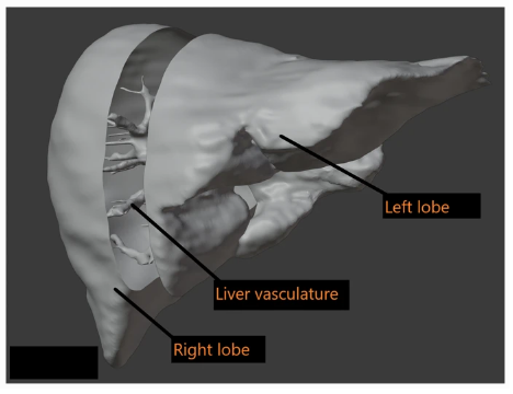
The liver was sectioned into 3 components digitally using Blender software to fit the 3D printer bed
A commercial Rostock Max V2 3D printer was used, with source image data converted via segmentation software, as well as open-sourced Slicer for ‘cropping the organ of interest.’ Structural supports were required for each 3D printed model, so post-processing work was involved.
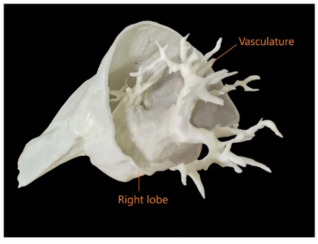
The right lobe of the liver was printed as 1 of 3 sections and joined to the vasculature

The outer renal cortex and inner calyces were separately printed as shells
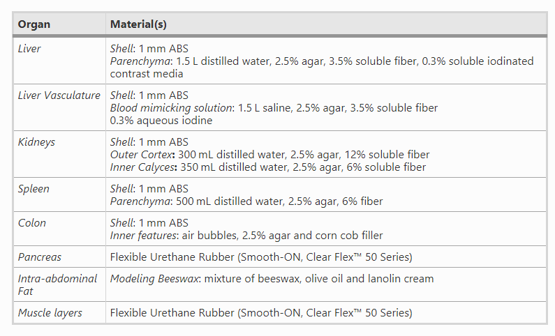
Each of the hollowed 3D printed cavities was filled out with attenuating material for greater accuracy. Central vasculatures were filled with contrast media/saline, and water-soluble agar filled the cavities.
“To fill the 3D printed organ shells with the agar, distilled water and fiber solution, a 250 mL syringe was inserted into a small opening in the organ shell,” stated the researchers.

A cross-sectional view of the internal cavities of the two halves of the kidney demonstrates filling with agar solution (light blue) for a radiological match
3D Printed organs and their components
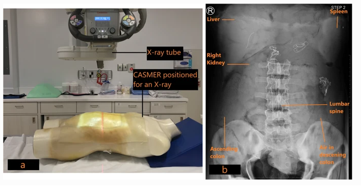
a: CASMER was positioned for an anteroposterior abdominal radiograph to determine radiological density. b: Anteroposterior X-ray of CASMER demonstrates the 3D printed organs and other structures as labeled
During the study, the researchers stressed that while structural accuracy was necessary, it was not as critical as radiological accuracy. Materials had to ‘closely mimic tissue with respect to radiological properties,’ and a thorough review was required by a radiologist. Image acquisition had to be excellent as well—and the researchers use the example of detailed internal liver vasculature as an example.
The researchers stated that during the project, both the small and large bowel were the most challenging to segment:
“The CT scan data that was available was suboptimal for segmentation and 3D printing of the bowel. Therefore, the decision was made to utilize an artistic rendering of the large and small bowel that could be more easily scaled to fit within the phantom cavity. Considerable editing of the shell was necessary to make a continuous hollow channel from the gastric sphincter all the way to the anus. Four threaded plugs were also created to allow access to the interior of the bowel for the purposes of adding radiopaque material to simulate obstructions and other material normally found in the digestive tract.”
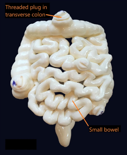
The 3D printed large colon and small bowel segments is demonstrated, with threaded plugs for internal access
“CASMER will be available for training medical radiation technology (MRT) students in cross sectional anatomy of the abdomen and for radiation dosimetry calculations,” concluded the researchers. “We will also explore 3D printing of pathologies within organs to facilitate training in performing image guided procedures.
3D printed models are helpful in a wide array of applications for medicine, allowing doctors to diagnose and treat conditions, educate patients, train medical students, and plan for surgeries too.

Intra-abdominal fat was mimicked with beeswax and formed a secure agent to house the removable 3D printed organs
Subscribe to Our Email Newsletter
Stay up-to-date on all the latest news from the 3D printing industry and receive information and offers from third party vendors.
Print Services
Upload your 3D Models and get them printed quickly and efficiently.


You May Also Like
APAC’s 3D Printing Capital Wave Is Bigger Than Venture Funding
By the usual measure, a tally of funding rounds, APAC’s additive manufacturing market had a quiet second quarter. The capital that has actually closed across the region comes to about...
HP Stock Jumps on 3D Printing Buzz
HP (NYSE: HPQ) had its best day in over a year this week, with shares jumping more than 7% on Tuesday. Interestingly, the move was quickly tied to 3D printing,...
3D Printing News Briefs, April 8, 2026: LiDAR Scanning, Vapor Smoothing, FDM Optimization, & More
We’ll kick off today’s 3D Printing News Briefs with some 3D scanning news from Artec 3D, and then move on to new America Makes Project Calls. Then, Raise3D and AMT...
3D Printing Market Hits $16B in 2025 as Growth Picks Up Again
The global 3D printing market reached $16 billion in 2025, growing just over 10% year over year, according to new data from Additive Manufacturing Research (AM Research). After a slower...


