
SWIFT: Uzel and Skylar-Scott are Paving the Way for the Future of Bioprinting


A few weeks ago, Mark Skylar-Scott and Sébastien Uzel, researchers working in Jennifer Lewis’ Lab at Harvard´s Wyss Institute for Biologically Inspired Engineering and John A. Paulson School of Engineering and Applied Sciences (SEAS), came up with a breakthrough new technique that could one day provide organ tissues for therapeutic use. The method, called SWIFT (sacrificial writing into functional tissue), allows 3D printing to focus on creating the vessels necessary to support a living tissue construct.
![]()

All organs need blood vessels to supply their cells with nutrients, but most lab-grown organoids lack a supportive vasculature. This is where the SWIFT method comes into play, 3D printing vascular channels into living tissues. Two weeks ago, 3DPrint.com went into some of the main details of the research, but now we have gone straight to the source and spoken with two of the co-first authors of the paper, which came out on September 6 in Science Advances, to understand the process behind the method, as well as the collaborative work shaping the future of Harvard’s bioengineering aspirations.
“Inspired by the 3D bioprinting techniques emerging from the Lewis lab and the community in general, Mark [Skylar-Scott] and I decided that is was time to tackle, head-on, the challenge of cell function and density, and tissue volume, which were keeping us from reaching organ manufacturing at therapeutic scale,” revealed Uzel. “Using patient-derived organoids or 3D cell spheroids as our building blocks appeared like a natural choice. They are cellularly dense and exhibit great functional and architectural similarities with the organs they are meant to mimic.”
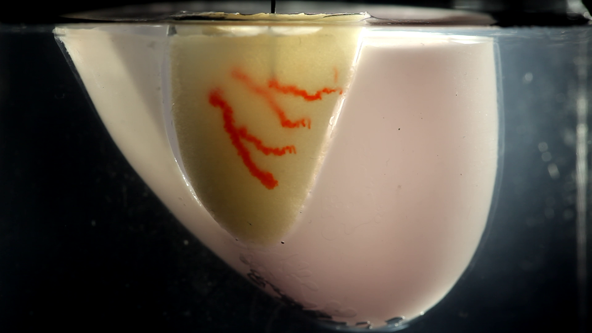
A branching network of channels of red, gelatin-based “ink” is 3D printed into a living cardiac tissue construct composed of millions of cells (yellow) using a thin nozzle to mimic organ vasculature.
Uzel went on to explain that “the idea of this SWIFT printing process really took shape when we speculated that once jammed into a dense slurry, those organoids would behave as predicted by the science of colloid suspensions and therefore could serve as a supporting living matrix for the free form templating of perfusable vessels. The rest was many months of testing and optimization!”
Both researchers and their colleagues found a way to pack living cells tightly enough together to replicate the density of the human body. Actually, they assembled hundreds of thousands of organ building blocks (OBBs) composed of patient-specific-induced pluripotent stem cell-derived organoids, which offer a pathway to achieving tissues with the requisite cellular density, microarchitecture, and function required. At the same time, they introduced vascular tunnels via embedded 3D bioprinting in between the OBBs to mimic blood vessels that are needed to deliver fluids, like nutrients and oxygen, that are vital to survival.
As an example, the group of researchers created a perfusable cardiac tissue that fuses and beats synchronously over a seven-day period. The SWIFT biomanufacturing method enables the rapid assembly of perfusable patient and organ-specific tissues at therapeutic scales. What is so novel about the new lab-grown heart tissue is that it beats, just like a normal human heart, and has an embedded network of the blood vessels that would be needed to survive if it was ever transplanted into a patient. It still needs to be tested before it can be used in humans, and their channels aren’t yet truly blood vessels, but if the innovation works for heart tissue, the experts expect SWIFT could also be used for other organs.
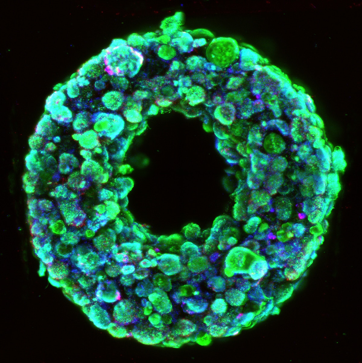
Living embryoid bodies surround a hollow vascular channel printed using the SWIFT method.
“We believe that this new technique addresses the technical roadblocks of cell density and manufacturing scalability. From a biology standpoint, making each building block more functional and performant, meaning being able to contract stronger in the context of cardiac tissues, for instance, is among the challenges that need to be overcome and will require gaining even more insights in pluripotent cell differentiation and how it can be recapitulated in vitro. We will also need to better emulate the multicellular and hierarchical complexity of the vessels as found in the human body,” proposed Uzel.
The researchers consider that on the manufacturing side of the process, the cost of reagents for scaling up cell culture and differentiation will have to be drastically reduced for de novo organ manufacturing to be a viable option looking into the long term.
When it comes to considering SWIFT as one of the main advances in the last few years towards bioprinting organs, Skylar-Scott claims “it would be presumptuous to say that SWIFT came out of a vacuum”.
“There have been many great works in this decade that have applied 3D printing to generate perfusable tissues, and our work builds on these efforts. What really does get us excited about SWIFT is how we have brought the matrix for embedded printing ‘to life’, and, by using organoids, we hope that SWIFT may serve as a bridge between the bottom-up self-assembly of developmental biology, and the top-down directed assembly of 3D printing,” Skylar-Scott asserted. “We can say, with reasonable certainty, that any successful engineering of a complex organ from scratch will require a combination of these two approaches.”
“The recent progress in the field of bioprinting has brought us a lot closer to the eventuality of 3D printed organs. The field is moving faster than we expected. Just five years ago, we were afraid to use “the big O word” [organs], but we are now, as a field, beginning to tentatively see a path forward,” he continued.
SWIFT is one of the projects at Harvard that could ultimately be used therapeutically to repair and replace human organs with lab-grown versions containing patients’ own cells. There is actually so much research at Wyss and SEAS, from scaling up tissue engineering to engineering miniature kidneys, it’s even one of the first places where researchers entirely 3D-printed an organ-on-a-chip with integrated sensing. Moreover, the creation of highly-organized multicellular biological tissues and organoids is structurally diverse and complex, so tissue manufacturing techniques require extreme precision, making us wonder what type of bioprinter the researchers are using.
According to Skylar-Scott, they “exclusively use custom made printers and extruders” in the lab, that “for the purposes of wacky experimentation, they offer the most versatility by far.” He also suggests that these printers are large and expensive, “but, for many processes, including SWIFT, we’re confident that it can be replicated with commercially available or open-source alternatives.”

As part of the SWIFT project evolution, collaborations are underway with Wyss Institute faculty members Christopher Chen, Professor of Biomedical Engineering and director of the Tissue Microfabrication Laboratory at Boston University and Sangeeta Bhatia, Professor at MIT’s Institute for Medical Engineering & Science (IMES) and Electrical Engineering & Computer Science (EECS), to implant these organ-specific tissues created by SWIFT into animal models and explore their host integration, as part of the 3D Organ Engineering Initiative, co-led by 3D printing pioneer and Wyss core faculty member, Jennifer Lewis, and Chen.
“We are currently working on rodent models for our initial in vivo phase. Along with perfecting our technique and improving the performance of printed tissues, we are investigating how small vascularized SWIFT-printed cardiac constructs integrate within the animal and connect to the existing blood stream. Once confident that the SWIFT tissues behave appropriately in small animals, the hope is to move to larger chunks of tissue to be tested on larger animals, in preparation for tests in humans in the long run,” revealed Uzel.
The collaborative work to make SWIFT a reality is a great example of integrating various disciplines and professionals into bioprinting projects.
“A process like SWIFT combines various expertise, from developmental biology to materials science or mechanical engineering. The strength of the lab is that it is built around great talents in all those disciplines. The Lewis lab is roughly divided into bioprinting and non-bioprinting work, but the two groups share technologies, techniques, and printing inks very frequently,” said Scott.
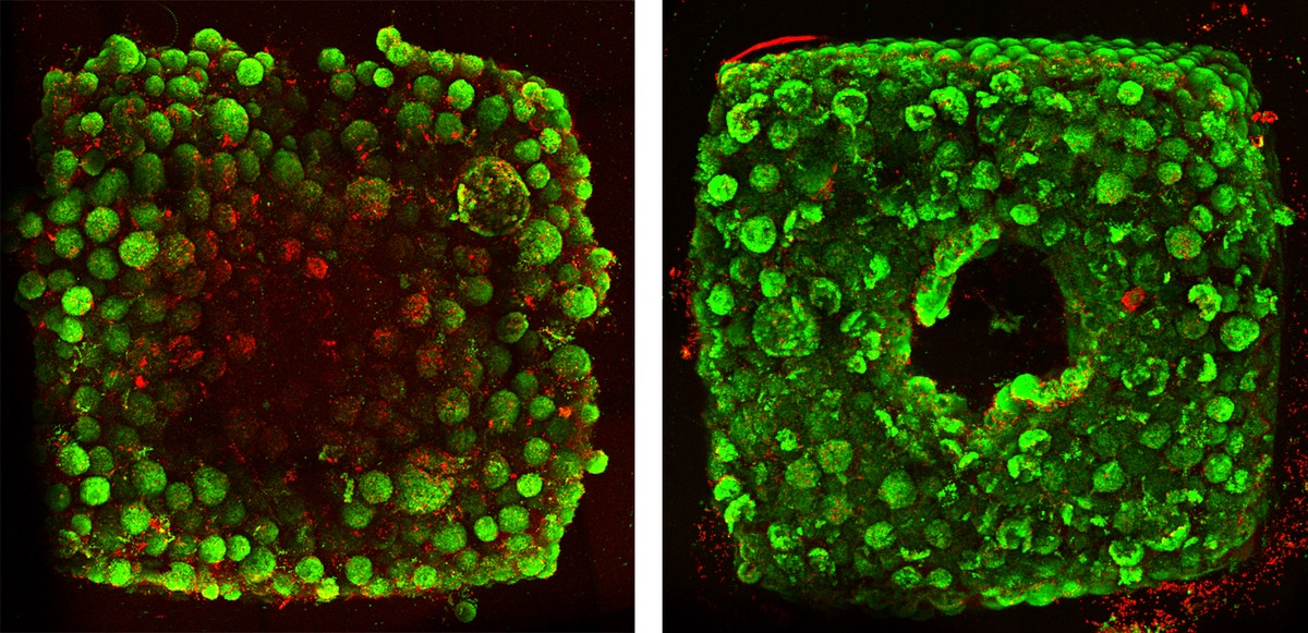
Tissues created without SWIFT-printed channels display cell death (red) in their cores after 12 hours of culture (left), while tissues with channels (right) have healthy cells.
He went on to explain that “it is unlikely that 3D printing can print all length-scales of an organ – from centimeter-scale ventricles to micrometer scale capillaries. So, we specifically designed the SWIFT process so that it can work with ‘organoids’ being built by the stem cell and developmental biology communities. By bridging the 3D printing and organoid fields, we believe there is a great potential for collaboration, and have already heard from researchers interested in using SWIFT to test scaling up their organoid systems. This interest has come from all sorts of specialists in different organs, including kidney, liver, heart, and brain.”
With so much going on, a typical day at the lab for Uzel and Skylar-Scott is not so typical. Although most of the daily tasks involve a combination of cell culture, printing ink formulation and characterization, CAD design and fabrication of printing and perfusion systems, tissue maintenance, imaging, and analysis. At busy times, Skylar-Scott says they could have upwards of four hours of work per day just to keep their cells fed, which has led to many long nights and weekends in the lab.
Similar to most academic labs, graduate students and postdocs all have two or three projects running in parallel.
“For SWIFT, we had to culture so many cells for a single print, that we were only running about one print per week. Since staring at cells doesn’t make them grow faster, it is often helpful to have a second project to focus on,” joked Skylar-Scott.
For example, they are currently working on new 3D printer hardware technology and focused on testing the SWIFT printed tissues in vivo so they can begin to test for additional function. All in a day’s work.
[Image Credit: Wyss Institute at Harvard University, John A. Paulson School of Engineering, Mark Skylar-Scott and Sébastien Uzel]Subscribe to Our Email Newsletter
Stay up-to-date on all the latest news from the 3D printing industry and receive information and offers from third party vendors.
Print Services
Upload your 3D Models and get them printed quickly and efficiently.


You May Also Like
Excellent Desktop Injection Molding, Made in Italy by Robot Factory
I was captivated when I saw my first Robot Factory 3D printer. The robust, precise machine was built to last. And this was in an era of very flimsy, disposable,...
Pogačar & Fairlight Cycles Show Us Low Cost 3D Printed Components for Bikes
There has been a lot going on in 3D printing for bicycles over the years. The most successful implementation so far is in bicycle seats. Carbon 3D printed seats are...
3D Printing News Briefs, June 18, 2026: Reseller, Relocation, Metal Space Powder, & More
We’ll start with business news in today’s 3D Printing News Briefs, as XJet appointed a value-added reseller in Germany, BIO INX is expanding its presence in the Italian market, and...
Researchers Combine AI and Bioprinting to Create Tiny Blood Vessel Networks
If 2026 has a theme in bioprinting, it may be blood vessels. Researchers can already print incredibly sophisticated tissues. The harder part is keeping those tissues alive. Without a network...


