NUS Researchers Created 3D Printed Training Models to Practice Wisdom Teeth Removal

During my senior year in high school, I completed that most loathsome of dental rituals – not braces, but having my wisdom teeth removed. My parents scheduled my oral surgery for the day after Christmas, which I claimed in all my teenage angst was cruel and unusual punishment, but really it was so I’d have more time to recover without having to take off extra days from school. The stories from that time are entertaining – I don’t know how my poor mother got a loopy, drugged-up 18-year-old back into the house by herself after the procedure, and once, I went out to the movies and instead of enjoying any popcorn, I spent the entire time alternating holding a giant cup of soda on each side of my sore face as a makeshift ice pack.
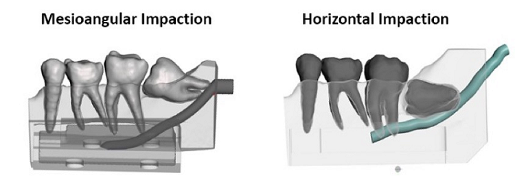
Impacted lower wisdom teeth are close to the inferior alveolar nerve, which can be injured during removal.
But I was lucky – my wisdom teeth were not impacted, so the oral surgeon didn’t have to cut into my gums to remove them, making both the procedure and the recovery easier.
Surgically removing superficially impacted wisdom teeth is a skill that dentists must learn. Common injuries that patients can suffer include excessive bone removal, burns to the lips and corner of the mouth, injuring adjacent teeth, and even injuring the inferior alveolar nerve, which is located directly beneath impacted wisdom teeth; this last can result in permanent lower lips and chin numbness. But, as always, practice makes perfect, and oral surgeons are less likely to accidentally injure patients as they continue to improve.
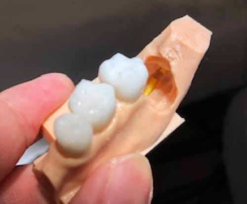
3D printed dental model with nerve – in this case there has been a nerve injury.
“The hardest thing to teach students is the actual ‘feel’ of the cutting instruments as it comes through many hours of practice. Unfortunately, in previous times, this learning happens after a complication that imparts wisdom to the learner but often times to the detriment of the patient,” said Dr. Raymond C.W. Wong from the National University of Singapore (NUS). “What if we can circumvent this process with the use of actual physical models that are realistic? The answer lies in 3D printing such models.”
Traditionally, students learn oral surgery methods by watching procedures being performed on patients, and then completing the procedure themselves. But, as those students could probably tell you, observing a procedure and performing it yourself are two very different things.

Students learning to perform oral surgery often use commercially available physical task trainers, but they’re limited due to cost, an unrealistic feel, and materials that crumble or melt when being drilled. We’ve seen 3D printing used in surgical training on many occasions, and a team of researchers from the NUS National University Centre for Oral Health, led by Dr. Wong, are doing just that in a new project funded by a grant from the National Additive Manufacturing Innovation Cluster (NAMIC).
Incorporating 3D printed surgical task trainers into the Faculty of Dentistry/National University Centre for Oral Health, NUS.
The team created a method for combining materials in order to create 3D printed impacted tooth models that include pulp, the pulp chamber, nearby nerves, and simulated periodontal ligaments.
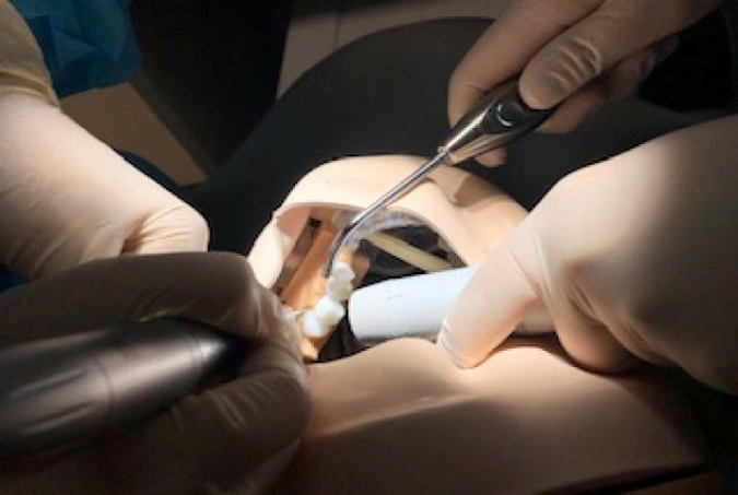
The material replicates the actual feel of bone and teeth being drilled for students, and can be printed, in varying degrees of difficulty and impaction, using scans from actual patients.
Then, the 3D printed model is mounted onto a mannequin head that features artificial cheeks, and the students can get to work, practicing proper instrument positioning so they won’t injure real patients, and repeating the procedure as many times as necessary.
By using these 3D printed wisdom teeth models, students who are practicing the procedure for the first time can enjoy increased confidence, and may be less nervous when the time comes to perform a real removal.
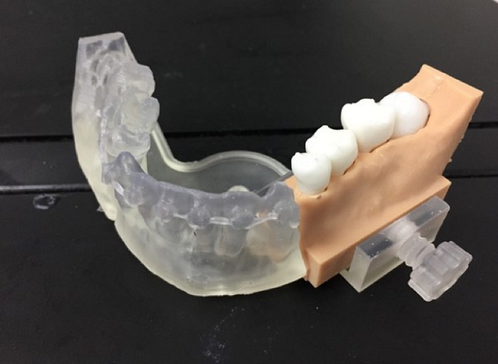
3D printed physical model-mandible base with replaceable cartridge containing the impacted tooth.
Discuss this story and other 3D printing topics at 3DPrintBoard.com or share your thoughts in the Facebook comments below.
[Images provided by Dr. Raymond Wong, NUS]Subscribe to Our Email Newsletter
Stay up-to-date on all the latest news from the 3D printing industry and receive information and offers from third party vendors.
Print Services
Upload your 3D Models and get them printed quickly and efficiently.


You May Also Like
Srini Kaza Discusses Strategically Scaling Align’s “Smile-Changing” 3D Printed Aligners
Align Technology‘s Invisalign is a revolutionary method to get you the smile you want through 3D printing. It is also a hugely popular process to go through, a $4 billion...
Analysis: Additive Manufacturing Strategies Spotlights Vertical Value Creation
A slowdown in the industry and particularly a tightening of access to capital following the additive manufacturing (AM) industry’s peak special purpose acquisition company (SPAC) phase in early 2021 is...
Department of Defense Awards PROTECT3D $1.3M for Custom 3D Printed Braces
Custom prosthetics company PROTECT3D has secured a $1.3M Department of Defense (DoD) Small Business Innovation Research contract to develop a platform for creating custom braces for soldiers. The PROTECT3D Platform...
AMS 2025 Highlights Big Changes in 3D Printing with Pivotal Speakers and Panels
2023 was filled with excitement around the potential mergers being pursued by industry stalwart Stratasys (Nasdaq: SSYS), leading to some of the most insightful conversations imaginable at Additive Manufacturing Strategies...