
What microCT Scanning Can Do for Additive Manufacturing


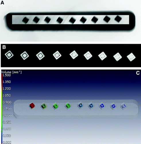
X-ray and CT of LPBF Ti6Al4V ELI test sample with designed internal cavities of varying dimensions. The contrasted X-ray image (A) reveals the presence of all the cavities, while CT allows much clearer 3D visualization and quantitative assessment as seen in a CT slice image (B) and 3D rendering (C). LPBF, laser powder bed fusion.
X-ray microcomputed tomography (microCT) has become widely used as a way to analyze and test additively manufactured parts, particularly for dimensional measurement and porosity analysis. In a paper entitled “X-Ray Microcomputed Tomography in Additive Manufacturing: A Review of the Current Technology and Applications,” a group of researchers takes a look at the various ways microCT has been used in additive manufacturing, and the benefits and limitations of each.
“X-ray microCT works on the principle of irradiating a sample with a beam of X-rays, measuring the subsequent absorption X-ray image, and repeatedly acquiring such images as the sample rotates,” the researchers explain. “The X-ray absorption (so-called projection) images represent views of the sample from many angles, providing internal detail due to the penetration of X-rays. The acquired images are then used in a mathematical reconstruction process to generate a volumetric data set. This volume comprises voxels (volumetric pixels) with the brightness of each pixel related to the X-ray density of the material it represents (X-ray density depends on physical density and atomic mass).”
One application discussed is porosity and defect analysis. Porosity refers to voids in a 3D printed part, caused by any of a variety of factors. MicroCT can be used to detect porosity in smaller 3D printed parts, but it can miss small pores in larger parts. Another application is volumetric density measurement. Additively manufactured parts are still regularly subjected to the Archimedes test to measure their volumetric density, but their are several problems with this method, according to the researchers:
-
Air bubbles can attach to the surface especially due to the rough and irregular surface, resulting in larger volume measurement and hence lower measured density.
-
Channels or flaws connected to the surface can be filled with water, resulting in a smaller volume measurement and hence a higher measured density.
-
Inclusions can potentially increase the measured mass and hence measured density.
-
Major flaws such as layer defects, keyhole or gas pores take very little volume (e.g., <0.01%) and hence do not make much difference to the measured density but can still be very important if they are clustered or layered, or have some nonrandom distribution.
-
A material density is assumed, which can be incorrect for alloys with varying compositions.

Example of porosity analysis in a small cylindrical sample (3 mm diameter) produced by LPBF with nonoptimal processing parameters, leading to total porosity of 4.5%. The porosity can be seen in an X-ray image (A) and visualized and quantified in different ways from CT data, shown in a 3D cropped view (B), CT slice image showing unmelted powder inside the pores (C), or a transparent view of the porosity analysis in 3D (D).
CT measurement can overcome the problems with bubbles and porosity or channels; the accuracy of the measurement is limited only by scan resolution.
Dimensional measurement is another application; in fact, microCT is the only method capable of measuring the dimensions of complex parts with internal surfaces and lattices. MicroCT can also monitor changes in parts, particularly after deformation. Another application is the measurement of surface roughness or topography; traditional tools can only measure the outer surfaces of parts while microCT can measure complex or interior surfaces.
The researchers also point to simulations as an application that has not yet been widely explored for microCT and additive manufacturing.
“Unlike simulations based on design geometry of parts, the actual parts, including defects, surface imperfections, and build errors, can be simulated, providing a theoretically more accurate prediction of the properties of the part,” they state. “This can be useful for two reasons: first, the actual effect of a defect on the resulting mechanical properties can be assessed, assisting to make pass/fail decisions on the use of a part; and second, the effect of defects can be studied and correlated with various mechanical tests on the same parts.”
Multiscale CT and fast scanning are discussed as ways to detect porosity in single parts in a cost-effective way. Powder analysis is another application, one that can detect pores in metal powders. While most work using microCT in additive manufacturing has focused on single materials, it is also possible to analyze multiple materials in a part, though the quality of the images may vary depending on the materials being investigated.
The researches point out some limitations of microCT, such as part size.
“When a part is too big, penetration of X-rays becomes an issue requiring high scan voltages, beam filtration, and resulting loss of quality in the images,” they explain. “This can often result in lack of detection capability on small pores and can lead to some edges of the part being less or more bright than others in the CT data, making a good surface model impossible, or very time-consuming, to correct using image processing methods. This is especially true of denser metals, and objects larger than 100 mm. The data can still be used for viewing for major defects but any more advanced analysis becomes much more challenging.”
This can be overcome by using higher voltage systems or lots of beam filtration. Advantages of microCT include the fact that it does not require a great deal of time invested, though more time is required for critical parts.
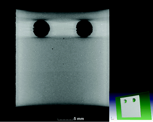
Isolated pores in a compact tension specimen, before the notch being machined into it (between the holes). Black spots indicate small isolated pores that can affect the crack growth in a fatigue test. Also visible are bright spots, which are impurities (inclusions), which may also act as stress concentration points and hence affect the fatigue crack growth behavior.
You can learn more detail about the many applications of microCT in the paper itself.
Authors of the paper include Anton du Plessis, Igor Yadroitsev, Ina Yadroitsava, and Stephan G. Le Roux.
Discuss this and other 3D printing topics at 3DPrintBoard.com or share your thoughts below.
Subscribe to Our Email Newsletter
Stay up-to-date on all the latest news from the 3D printing industry and receive information and offers from third party vendors.
Print Services
Upload your 3D Models and get them printed quickly and efficiently.


You May Also Like
Australia’s AMCRC Funds Titanium 3D Printing R&D
In terms of the global economy’s presently existing state, there is no realistic path to economic resilience that doesn’t start with critical minerals security. This is a problem for pretty...
3DPOD 305: Automating AM with Grenzebach’s Oliver Elbert
Oliver Elbert‘s over ten years in additive manufacturing have been spent automating LPBF. For large, high-volume, or critical parts, Grenzebach has provided custom automation solutions. Depowdering, powder handling, sieving, heat...
AMPulse Asia: Chinese IPOs, Defense Deals, and Dental 3D Printing Lead APAC Roundup
The second half of June brought a wave of additive manufacturing activity across China, Japan, South Korea, India, and Australia. From Chinese IPOs and funding rounds to defense, aerospace, construction,...
Austal, Curtin University and AMCRC Work on R&D Together
Australia’s Additive Manufacturing Cooperative Research Centre (AMCRC) works with 70 industry partners to deliver collaborative R&D projects. They also work on workforce development and technology transfer. It’s kind of analogous...


