
For Hope & Faith: The Power of 3D Printing Helps Separate Conjoined Twins

 Often, we talk about many new innovations and processes in manufacturing that would simply not be possible without the power of 3D printing behind them.
From automotive to aerospace, many companies are embracing this technology, and are rewarded with better, stronger components that can be produced more rapidly and affordably. However, whether we are producing better cars, faster planes that are easier to maintain, or heading into space with greater self-sustainability and a more attractive bottom line–the reality is that we are discussing cold, hard, inanimate materials.
Looking into the medical industry, the story of 3D printing grows much warmer. There, we are often dealing with the human heart and spirit, and often the fight for a better life, or quite simply–life itself. Traditional and new technologies are able to complement one another, working together, as medical professionals are able to take items like CT scans and MRIs and convert them into a range of 3D models for devices, implants, and more.

Dr. Rajesh Krishnamurthy, chief of radiology research and cardiac imaging at Texas Children’s Hospital.
One of the greatest benefits being seen from the use of 3D medical models is that doctors are using them for diagnosis, educating patients and medical students, prepping for surgery–and using them in the operating room for navigating the most complex parts of new surgeries. While this is tremendous for the outcomes of patients, it’s an incredible tool for helping surgeons to take on–and train for–procedures that may not have been previously possible, as well as allowing for better efficiency, and often resulting in less time with patients under anesthesia. We’ve followed countless procedures making a perfect example of this, from helping with tumor removals to that of surgeons saving a female patient’s kidney recently.
Complex is certainly an understatement when it came to a recent surgery separating conjoined twins. Made possible with the help of 3D printing, the details were released in a recent study presented at the annual meeting of the Radiological Society of North America (RSNA).
Conjoined twins are rare (one in every 200,000 live births in the US), as are the consequent and extremely difficult surgeries for separation; however, success has become much better in the past 65 years.
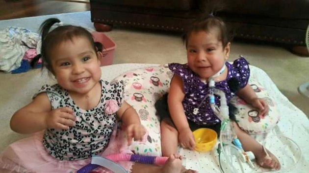
[Image credit: RSNA/Texas Children’s Hospital]
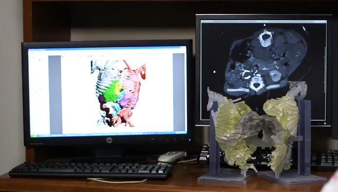
“This case was unique in the extent of fusion,” said lead author Dr. Rajesh Krishnamurthy, chief of radiology research and cardiac imaging at Texas Children’s Hospital, in a press release. “It was one of the most complex separations ever for conjoined twins. The CT scans showed that the babies’ hearts were in the same cavity but were not fused. Also, we detected a plane of separation of the liver that the surgeons would be able to use.”
Helpful for much more than just showing positioning of the babies’ vital organs, doctors were also able to see the CTs converted into complex, color coded (corresponding to each girl) 3D models which then comprehensively detailed the skeletons, supports, livers, and blood vessels of the two girls. This incredible visual display helped them to make a plan for the surgery in February, which lasted for 26 hours.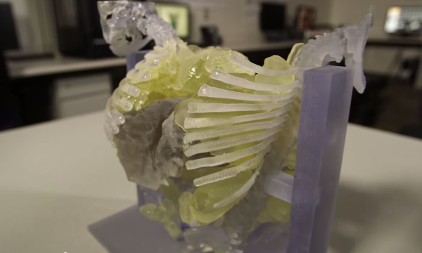
“The surgeons found the landmarks for the liver, hearts and pelvic organs just as we had described,” Dr. Krishnamurthy said. “The concordance was almost perfect.”
Including a dozen surgeons and a vast number of other medical personnel, the procedure was a success, and will most likely serve as a technique in the future for similar surgeries.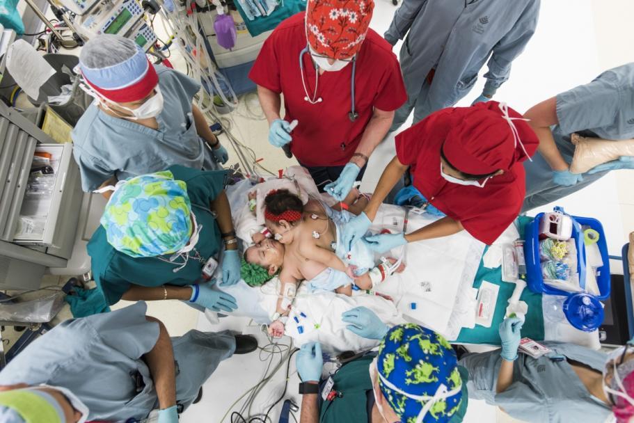
One bonus for making these 3D models, while they help surgeons to do things perhaps not previously possible, is that they are enormously helpful also in explaining and outlining to patients (and their families) exactly what will be happening in the procedure, where, and why. This was certainly the case for the parents of the twins, who were more comfortable after being able to understand the procedure more fully due to the help of the 3D model.
The twins were in the hospital for several months following the surgery, with Hope being released in May, and Faith following suit a month later. They are both at home and doing well. Discuss this story in the 3D Printing and Conjoined Twins forum on 3DPB.com.
 [Source: Medical Daily]
Subscribe to Our Email Newsletter
Stay up-to-date on all the latest news from the 3D printing industry and receive information and offers from third party vendors.
Print Services
Upload your 3D Models and get them printed quickly and efficiently.


You May Also Like
3D Printing News Briefs, March 28, 2026: TCT Asia, Distribution Agreement, FDA Clearance
We’re starting 3D Printing News Briefs this weekend with some news out of TCT Asia, and then moving on to a metal AM distribution agreement between MULTISTATION and WAAM3D. We’ll...
The Magic of AMUG as Reported by a First-Time Attendee
There’s a special kind of magic about AMUG. I’ve heard about it for years, but never experienced it myself until last week. It’s different than what you see at some...
3D Printing News Briefs, March 26, 2026: AMUK, IP Dispute, Asbestos, & More
We’re kicking off today’s 3D Printing News Briefs with an America Makes Project Call, and then moving on to additive manufacturing in the UK. Then we’ve got some legal news...
At TCT Asia 2026, China’s AM Industry Looked Ready for Scale: Part 1
Walk into TCT Asia 2026, and the first impression is density. More than 55,000 square meters across Halls 7.1 and 8.1 at Shanghai’s National Exhibition and Convention Center, over 550...