Tel Aviv University: Researchers 3D Print Cardiac Patches & Cellularized Hearts

 Researchers at Tel Aviv University continue to try to meet the ongoing challenges in cardiac tissue engineering. In ‘3D Printing of Personalized Thick and Perfusable Cardiac Patches and Hearts,’ authors Nadav Noor, Assaf Shapira, Reuven Edri, Idan Gal, Lior Wertheim, and Tal Dvir outline the steps they took to match technology with tissue.
Cardiovascular disease is the leading killer of patients in the US, and organ donor and transplantation processes can still mean a long wait for those suffering from heart failure. Here, the authors demonstrate the need for alternative ways to treat the infarcted (usually referring to clogging of one of more arteries) heart. And while tissue engineering has pointed the way to freeing many patients from terrible physical suffering and organ donor waiting lists, creating the necessary scaffolds with true biocompatibility has presented obstacles.
The authors have created an engineered cardiac patch meant to be transplanted directly onto the patient’s heart, integrating into the ‘host,’ with excess biomaterials degrading over time. This leaves the cardiac patch, full of live, healthy tissue, regenerating a previously defective heart. Because there is always the threat of rejection when implanting anything into the body though, the authors emphasize the need for appropriate materials:
“Most ideally, the biomaterial should possess biochemical, mechanical, and topographical properties similar to those of native tissues,” state the researchers. “Decellularized tissue‐based scaffolds from different sources meet most of these requirements. However, to ensure minimal response of the immune system, completely autologous materials are preferred.”
The researchers were able to create patient-specific cardiac patches in their recent study, extracting fatty tissue from cardiac patients—and then separating cellular and a-cellular materials.
“While the cells were reprogrammed to become pluripotent stem cells, the extra‐cellular matrix (ECM) was processed into a personalized hydrogel,” stated the researchers. “Following mixture of the cells and the hydrogel, the cells were efficiently differentiated to cardiac cells to create patient‐specific, immunocompatible cardiac patches.”
In using the patient-specific hydrogel as bioink, the researchers were able to create patches, but ultimately, they were also able to 3D print comprehensive tissue structures that include whole hearts.
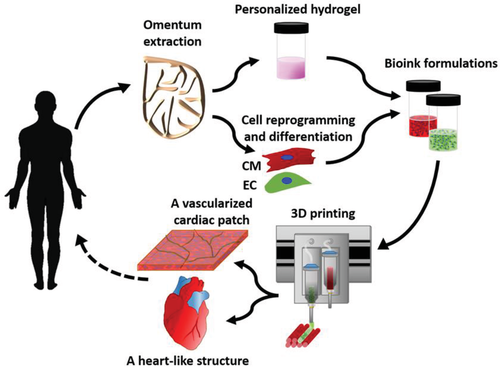
An omentum tissue is extracted from the patient and while the cells are separated from the matrix, the latter is processed into a personalized thermoresponsive hydrogel. The cells are reprogrammed to become pluripotent and are then differentiated to cardiomyocytes and endothelial cells, followed by encapsulation within the hydrogel to generate the bioinks used for printing. The bioinks are then printed to engineer vascularized patches and complex cellularized structures. The resulting autologous engineered tissue can be transplanted back into the patient, to repair or replace injured/diseased organs with low risk of rejection.
The authors used two different models in their study, with one serving as proof-of-concept, with pluripotent stem cells (iPSCs)‐derived cardiomyocytes (CMs) and endothelial cells (ECs). The other model relied on:
- Rat neonatal CMs
- Human umbilical vein endothelial cells (HUVECs)
- Lumen‐supporting fibroblasts
One bioink, laden with cardiac cells, printed parenchymal tissue, while the other extruded cells for forming blood vessels. The researchers were successful in 3D printing the patient-specific cardiac patches but found when a higher degree of complexity was necessary for fabrication of organs or other tissues, the hydrogels were not strong enough. They created a new process for organs and more complex tissues where they could print in a free-form manner and cure structures at varying temperatures; they were able to overcome previous challenges and 3D print accurate, personalized structures.
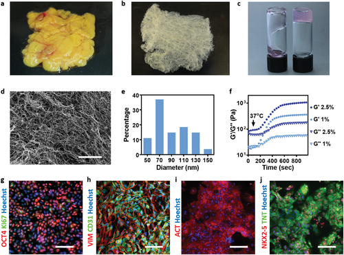
Bioinks characterization. A human omentum a) before and b) after decellularization. c) A personalized hydrogel at room temperature (left) and after gelation at 37 °C (right). d) A SEM image of the personalized hydrogel ultrastructural morphology, and e) a histogram of the fibers diameter. f) Rheology measurements of 1% w/v and 2.5% w/v omentum hydrogels, showing the gelation process upon incubation at 37 °C. g) Stromal cells originated from human omental tissues were reprogrammed to become pluripotent stem cells (red: OCT4, green: Ki67 and blue: nuclei). h) Differentiation to ECs as determined by CD31 (green) and vimentin staining (red). Differentiation to cardiac lineage: i) staining for sarcomeric actinin (red), j) staining for NKX2‐5 (red), and TNNT2 (green). Scale bars: (e) = 10 µm, (g,i,j) = 50 µm, (h) = 25 µm.
This study carries substantial weight, considering the researchers were able to create cellularized hearts with ‘natural architectures.’ This furthers the potential for cardiac transplants after heart failure, along with encouraging the process for drug screening. The authors point out that more long-terms studies and research with animal models are necessary.
“Although 3D printing is considered a promising approach for engineering whole organs, several challenges still remain,” conclude the researchers. “These include efficient expansion of iPSCs to obtain the high cell number required for engineering a large, functioning organ. Additionally, new bioengineering approaches are needed to provide long‐term cultivation of the organs and efficient mass transfer, while supplying biochemical and physical cues for maturation.”
“The printed blood vessel network demonstrated in this study is still limited. To address this challenge, strategies to image the entire blood vessels of the heart and to incorporate them in the blueprint of the organ are required. Finally, advanced technologies to precisely print these small‐diameter blood vessels within thick structures should be developed.”
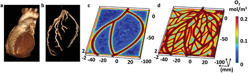
Imaging of the heart and patch modeling. CT image of a) a human heart and b) left ventricle coronary arteries. c) A model of oxygen concentration profile in an engineered patch. d) Replanning of the model showed better oxygen diffusion, sufficient to support cell viability.
Without good heart health, it is very difficult to survive. Responsible for transporting nutrients, oxygen, and more to cells populating the human body, the heart also removes waste like carbon dioxide and more. 3D printing is assisting scientists and doctors in researching and treating a variety of different diseases and conditions, whether they are using 3D printed metamaterials for fabricating heart valves, creating better cardiac catheters, or experimenting with new types of phantoms.
What do you think of this news? Let us know your thoughts! Join the discussion of this and other 3D printing topics at 3DPrintBoard.com.
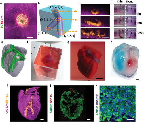
Printing thick vascularized tissues. a) A top view of a lumen entrance (CD31; green) in a thick cardiac tissue (actinin; pink). b) A model of a tripod blood vessel within a thick engineered cardiac tissue (coordinates in mm), and c) the corresponding lumens in each indicated section of the printed structure. d) Tissue perfusion visualized from dual viewpoints. e–k) A printed small‐scaled, cellularized, human heart. e) The human heart CAD model. f,g) A printed heart within a support bath. h) After extraction, the left and right ventricles were injected with red and blue dyes, respectively, in order to demonstrate hollow chambers and the septum in‐between them. i) 3D confocal image of the printed heart (CMs in pink, ECs in orange). j,k) Cross‐sections of the heart immunostained against sarcomeric actinin (green). Scale bars: (a,c,h, i,j) = 1 mm, (g) = 0.5 cm, (k) = 50 µm.
Subscribe to Our Email Newsletter
Stay up-to-date on all the latest news from the 3D printing industry and receive information and offers from third party vendors.
You May Also Like
Changing the Landscape: 1Print Co-Founder Adam Friedman on His Unique Approach to 3D Printed Construction
Additive construction (AC) is much more versatile than it seems, at first: as natural as it is to focus on the exciting prospect of automated home construction, there’s far more...
US Army Corps of Engineers’ Megan Kreiger on the State of Construction 3D Printing
Despite last year’s gloomy reports about the financial state of the additive manufacturing (AM) industry, there’s no doubt that we’re actually witnessing the birth of a sector rather than its...
3D Printing Webinar and Event Roundup: April 21, 2024
It’s another busy week of webinars and events, starting with Hannover Messe in Germany and continuing with Metalcasting Congress, Chinaplas, TechBlick’s Innovation Festival, and more. Stratasys continues its advanced training...
Profiling a Construction 3D Printing Pioneer: US Army Corps of Engineers’ Megan Kreiger
The world of construction 3D printing is still so new that the true experts can probably be counted on two hands. Among them is Megan Kreiger, Portfolio Manager of Additive...