

3D Printing Allows For Human Embryonic Stem Cell Breakthrough – Cell Organization


Human embryonic stem cells (HESC), for years have been the center of controversy. This is mainly due to moral issues, creating a dilemma in determining where the fine line between human life, and a simple human cell should 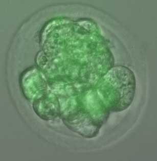 reside. Should HESCs have the same basic moral status as a human being? This is a question I am not qualified to answer, nor do I even want to consider at this point in time.
Scientists have been looking to get around this moral dilemma for years, coming up with a variety of methods which seem to be more humane than destroying an actual embryo to obtain the cells. With this said, obtaining HESCs is not the main problem for researchers, who have found it extremely difficult to reliably stimulate the cells, in order to form a specific type of human tissue.
The most amazing characteristic of an embryonic stem cell is the fact that it can turn into any of the 220 different types of cells within the human body. These 220 cells are derived from three main primary germ layers of cells which include the ectoderm, endoderm, and mesoderm. When researchers try and grow these cells in a petri dish, they have not been able to differentiate the areas in which each of the three germ layers are grown. Unlike within the human body, where chemical signals are sent to the HESC’s, via the surrounding tissue, telling them where to form, when allowing for HESC’s to grow in a lab, researchers find that the cells do not separate in the proper orientation.
To compensate for this lack of chemical signals, many researchers have tried creating their own signals with various chemicals found in their labs, but have been unsuccessful in trying to coax the cells to separate in the correct orientation.
In a recent paper published on Nature.com, researchers led by Ali Brivanlou, Robert and Harriet Heilbrunn, from the Laboratory of Stem Cell Biology and Molecular Embryology at Rockefeller University took an entirely different approach. Instead of relying on chemical signals to spur on the separation of the three different germ layers, they instead turned to geometry, with the help of 3D Printing.
Various types of cells differentiated by color
The researchers used 3D printed molds which were created out of a silicone-based elastomer called Polydimethylsiloxane (PDMS). By using 3D printing they were able to control the specific depth, shape, and diameter of each of the molds, meaning they could determine the exact size and shape of each HESC colony. They also found that the distribution of the cells within the molds were extremely uniform, meaning that they had more control over each colony within each mold. They then introduced various different stimuli into the equation, allowing them to determine which type of cells each mold would grow. The 3D printed molds allowed the researchers to make sure each colony of cells were separated from the other colonies. For the very first time they were not only able to coax the cells into differentiating themselves from one another, but also control the exact locations that each individual cell colony would form.
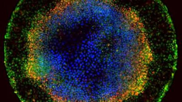
Various germ layers separated
“At the fundamental level, what we have developed is a new model to explore how human embryonic stem cells first differentiate into separate populations with a very reproducible spatial order just as in an embryo,” said Aryeh Warmflash, a postdoc who worked on this particular research. “We can now follow individual cells in real time in order to find out what makes them specialize, and we can begin to ask questions about the underlying genetics of this process. These cells have a powerful intrinsic tendency to form patterns as they develop. Varying the geometry of the colonies may turn out to be an important tool that can be used to guide stem cells to form specific cell types or tissues.”
The method used by these researchers could prove to be a major step towards future stem cell therapies, and even the regrowth of injured or lost human tissue. Let us know your opinion on this amazing research in the 3D printed stem cell mold forum thread on 3DPB.com.
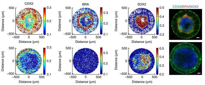
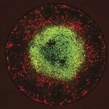
Diagram showing the separated germ layers achieved.
Subscribe to Our Email Newsletter
Stay up-to-date on all the latest news from the 3D printing industry and receive information and offers from third party vendors.
Print Services
Upload your 3D Models and get them printed quickly and efficiently.


You May Also Like
NASA Selects Relativity Space for Mars Science Mission
NASA has selected Relativity Space as its commercial partner for a new Mars science mission scheduled for launch in 2028, giving another boost to one of the most well-known additive...
AM Asia Watch: China’s 3D Printing Boom Is Creating a New Class of Micro-Manufacturers
China’s additive manufacturing (AM) industry has spent years trying to reduce its reliance on foreign technology. In polymer 3D printing, domestic companies have already become major players. In metal AM,...
Excellent Desktop Injection Molding, Made in Italy by Robot Factory
I was captivated when I saw my first Robot Factory 3D printer. The robust, precise machine was built to last. And this was in an era of very flimsy, disposable,...
Divergent Declares that German 3D Printers are Superior, And Plans Massive LPBF Expansion
Divergent has announced a new version of its Laser Powder Bed Fusion (LPBF) printer and a new site. The company aims to do nothing short of “further accelerating its mission...


