Boston’s Additive Edge: Inside Harvard’s Lewis Lab and the Bioprinting Blueprint, Part I

At Harvard’s School of Engineering and Applied Sciences (SEAS) and the Wyss Institute at Harvard, Jennifer Lewis’s lab is at the intersection of biology and engineering. Part workshop, part incubator for the future of medicine, here stem cells are coaxed into forming tissues, custom-built printers lay down living structures, and the ultimate ambition is creating organs that can heal, replace, or even save human lives.
Among the team leading this charge is Paul Stankey, a doctoral researcher in his fifth year, who has dedicated his training to one of bioprinting’s hardest problems: designing blood vessels that can keep engineered tissues alive.
I met with Stankey this summer at a Harvard lab in Cambridge, MA, where Lewis’s team is reimagining how bioprinting can move from research to the clinic. With a background that spans biomedical, mechanical, and even what he calls “a dose of electrical engineering,” Stankey described his role as pretty straightforward:
“Really, I’m a manufacturing engineer. That’s how I view everything I do. I’m making techniques to get developments out to patients as fast as possible.”
That focus runs through the entire Lewis Lab. Lewis, a pioneer in 3D bioprinting, has assembled a team that works across the boundaries between biology, engineering, medicine, and manufacturing. Her students are not only aiming for academic breakthroughs, but also for practical tools that could transform patient care.

Engineering Sciences Laboratory at Harvard. Image courtesy of 3DPrint.com.
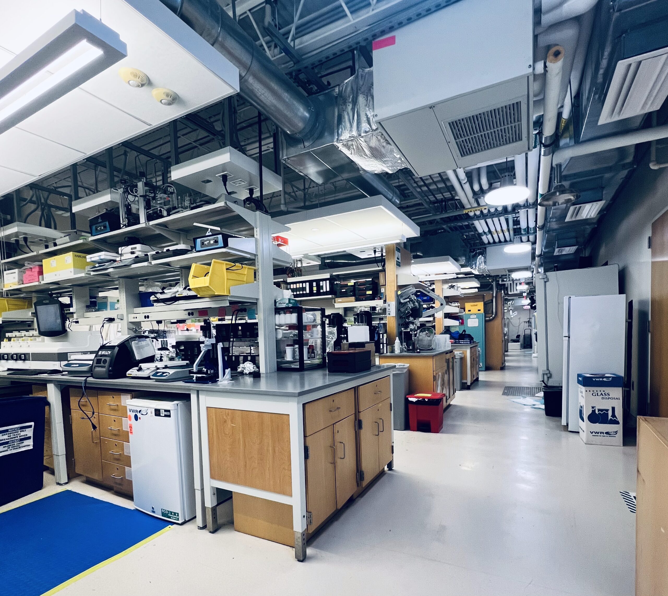
A Lab with No Limits
The lab is organized almost like a pipeline for organ-making, Stankey explained. The process begins with growing vast numbers of stem cells, then assembling them into tissue building blocks, and finally culturing them into more complex, organ-specific structures. The space itself reflects that sequence—stem cell production at the front, large bioprinters in the back, and materials processing in a separate lab downstairs.
“We have all the toys you could ever possibly want. Jennifer makes sure we’re well supplied. She never wants our work to be limited by resources — we’re only bound by physics, which is a fun place to be.”
This also shows up in the day-to-day workflow. Stankey pointed out that a researcher can literally print an ink, take it off the printer, and walk it six feet over to a tester to see results the same day.
“That’s one of my favorite parts of the lab,” he said. “You fail, but you know why you failed — and then you can try again right away.” Even the printers carry personality: some of the biggest machines are nicknamed “Robo Mama” and “Robo Daddy.”
That philosophy is the backbone of the workflow: a single bioreactor can generate up to two billion cells, enough material to produce around 40 mouse kidneys. Scaling those numbers up, Stankey explained, is how the team begins to think about moving from small-scale models to tissues that could one day help patients.
Those mouse kidneys don’t just stay in Cambridge. The lab grows and prints them in Cambridge, then someone drives them across town to Massachusetts General Hospital’s Charlestown research facility, where animal implantations are performed under strict regulation.
“We’ll sit in the operating room and watch a surgeon implant them,” Stankey said. “You see how they fail, sometimes in a split second, sometimes four hours later. That’s the information we need to make the next one better.”
Their current record? A printed kidney that stayed alive for five hours. Within a year, he said they “hope to be trucking mouse-sized human kidneys to MGH once a week.”
These kidneys are built from human induced pluripotent stem cells and tested in immunocompromised mice to avoid rejection. For Stankey and the team, it’s a crucial step: animal models provide the data needed to improve designs, while pointing toward the larger vision. If the same techniques are eventually matched with a patient’s own stem cells, those cells could be expanded, differentiated, and used to print tissues uniquely their own, reducing the risk of rejection and opening the door to personalized organ repair.

Four port device for SWIFT printing used to print mouse-sized kidneys. Image courtesy of 3DPrint.com.
Printing Blood Vessels That Work
“The hardest part of making large tissues isn’t just putting down cells. Creating a solid mass would be straightforward if you had enough of them. The real challenge comes when you need to 3D print the blood vessels that keep those tissues alive and functioning. Without vessels, thick tissues starve before they have a chance to work. That’s where our research has broken new ground.”
The lab first tackled this problem in 2019 with the development of SWIFT (Sacrificial Writing Into Functional Tissue). This method used a sacrificial ink to carve out hollow channels inside living cell matrices, which looks like a cluster or blob of cells. Those channels could then be lined with vascular cells, creating space for blood to flow. It was a breakthrough because, for the first time, scientists could build thick, “living tissues that didn’t suffocate in the lab.”
Then in 2024, Stankey co-authored a paper introducing co-SWIFT (or coaxial SWIFT), a method for printing multilayer vascular structures within living cardiac tissue.
“We’re essentially doing the same thing the body does,” he said. “We’re trying to build vessels at different scales. In the larger vessels — the ones that take all the blood pressure — you need a vessel wall. So we created a core-shell architecture, where the shell is smooth muscle and the lumen can be seeded with endothelial cells. You get a tri-layered structure, just like in your body.”
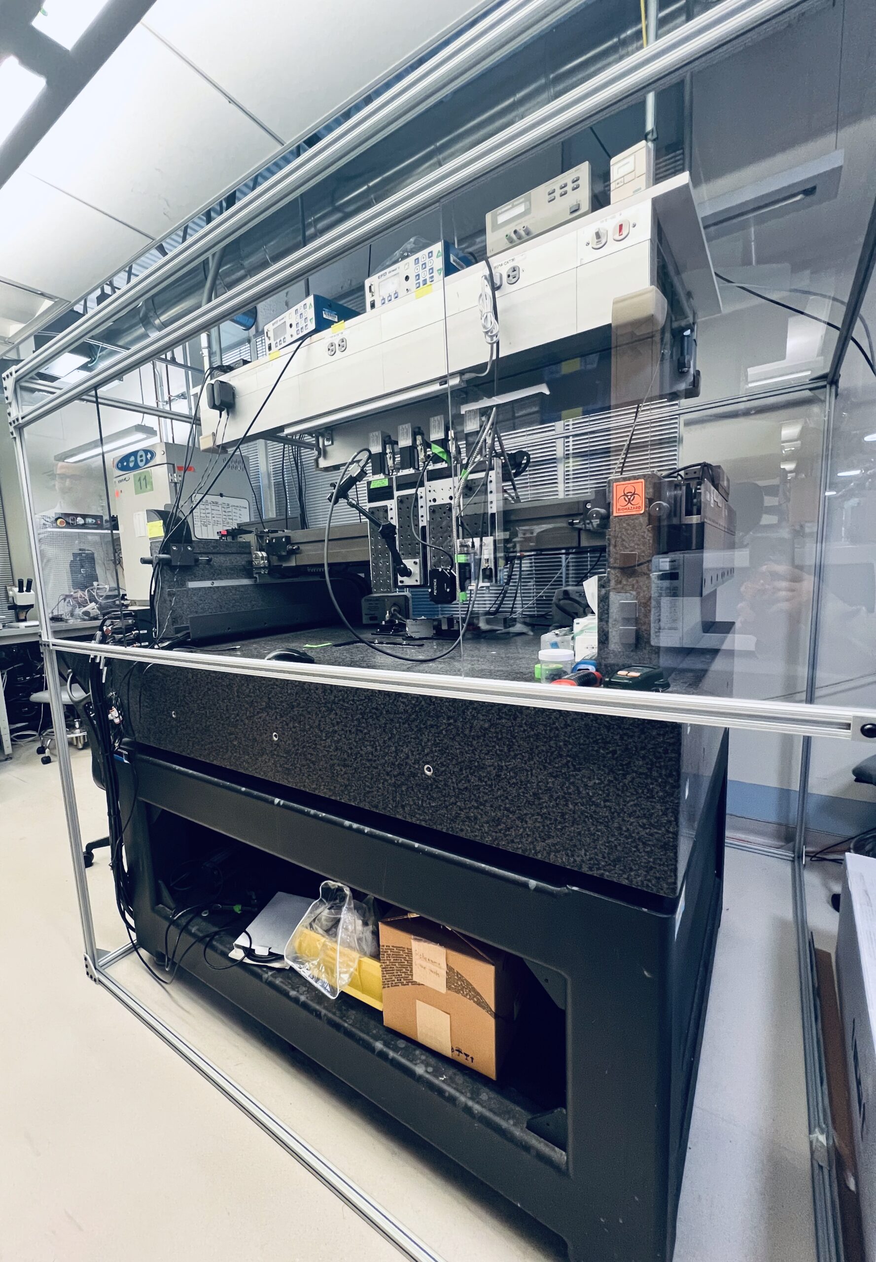
A multi-axis bioprinter at the back of Jennifer Lewis’ Lab, a custom machine developed in-house, first pioneered by Jennifer Lewis and her then-postdoc Mark Skylar-Scott. Today, it anchors much of the lab’s efforts in printing complex, living tissues. Image courtesy of 3DPrint.com.
What made co-SWIFT truly novel, Stankey pointed out, was learning how to print branching vascular networks.
“That was the leap. Not just one tube, but a system that splits and connects, the way your own vasculature does.”
The results were striking. The printed cardiac tissues not only developed vessels but also began beating synchronously and even responded to heart drugs the way native tissue does. “It was, in every sense, living,” Stankey said.
But he stresseed that “translation will be gradual. The first thing you’ll see in the clinic won’t be a full organ. It’ll be bits and pieces — patches, vascular grafts, those types of structures.”
One effort is through CellMet, a multi-institution consortium focused on cardiac patches. “If someone has a heart attack and part of their heart dies, a surgeon could place a patch on the outside. Right now, people get mechanical pumps, and those have all sorts of problems. But if you make a patch from a patient’s own cells, their immune system can help it integrate. That’s the biologically inspired part of engineering that guides how we design these engineered constructs.”
Jennifer Lewis’ Lab at Harvard’s SEAS. Image courtesy of 3DPrint.com.
Medicine, Nature, and Design
So, should a printed organ replicate its natural form, or could it be simplified?
“There’s a question in the field,” Stankey explained. “If you make a kidney, should it look like a kidney? Or should it look like a square? It’s easier to make squares than kidney shapes. But what we keep finding is that the body is really smart. Things look the way they do for a reason, after millions of years of evolution. Usually, after you try something different, you realize, ‘oh, we should’ve made a kidney.’”
Respect for nature is a big part of the lab’s identity. The Wyss Institute is, after all, dedicated to “biologically inspired engineering.” For Stankey, it’s a guiding principle, and it circles back to the heart patch he mentioned earlier.
“If I’m trying to treat a heart attack, maybe I don’t want to replace the whole heart. Maybe I just need a patch. It’ll be cheaper, faster to get to patients, and less invasive. That’s the kind of thinking that keeps us moving.”
He noted that such a patch might not even require open-heart surgery. While placing it through a vein, like in current ablation procedures, would be challenging, the hope is that the surgery could be minimally invasive.
“That’s why collaborating with clinicians is so important. We have to design in a way that works in the real world, not just in the lab. And having hospitals and surgeons right next door at Harvard and MGH makes that collaboration possible. It lets us see how these therapies could actually be delivered.”
The science is definitely bold, but the questions are bigger: How do you scale this up? How do you get from a mouse-sized kidney to a human heart or kidney? And most importantly, how do you deliver it safely to patients? Those challenges will be the focus of our next story, as the Lewis Lab works to bring these ideas closer to patients.
Subscribe to Our Email Newsletter
Stay up-to-date on all the latest news from the 3D printing industry and receive information and offers from third party vendors.
Print Services
Upload your 3D Models and get them printed quickly and efficiently.


You May Also Like
Cybersecurity: A Necessity in a Maturing Additive Manufacturing Industry
Additive Manufacturing is no longer an experimental technology operating on the margins of industrial production. Over the past decade, it has become an integral part of how OEMs design, produce,...
How Metal Additive Manufacturing Is Reshaping the Future of Aerospace and Defense Engineering
Additive manufacturing (AM) is steadily changing the way we think about producing metal parts for aircraft. Whilst aerospace and defense companies have been using metal AM for over twenty years,...
The AM Adoption Problem No One Models
Through its short history, Additive Manufacturing has been presented to the investment community as a classic high growth technology story. Manufacturing is a multi-trillion-dollar market. Even a small percentage looks...
The Business of Customized Sports Equipment: How 3D Printing Is Changing Athletic Gear
For years, 3D printing in sports has been linked mostly with prototypes, concept shoes, and one-off experiments for elite athletes. Helmets, cleats, and footwear midsoles often took over the headlines,...