
Diabetes Treatment Gets Sci-Fi Upgrade with 3D Printed Microdevice Eye Implant


In a revolutionary step forward, researchers have 3D printed a biohybrid microstructure designed specifically for diabetes treatment. This innovation, stemming from a collaboration between the KTH Royal Institute of Technology and Karolinska Institutet, introduces a medical microdevice intended for the human eye, providing a fresh road for cell-based medical treatments for the rising global population affected by diabetes type 1 and 2. To achieve this, the team utilized Nanoscribe‘s 3D printing technology and materials.
 The chip with insulin-producing cells. Image courtesy of KTH.
Think of it like a tiny 3D printed structure made of living cells, so small that it can fit inside the front part of the eye. By placing it there, doctors can keep a close watch on how it’s helping treat diabetes. This new invention could be a big step in helping people with diabetes in the future.
A rapidly growing global ailment, diabetes has been labeled as the world’s fastest-expanding public disease. Data from different institutions confirms the scale of this health crisis. The Swedish Diabetes Association reports a staggering 425 million people affected. At the same time, the World Health Organization (WHO) indicates that about 422 million people worldwide have diabetes. The majority of these individuals live in low-and middle-income countries. The International Diabetes Federation (IDF) further estimated that diabetes caused about 4.2 million deaths globally in 2019. This total includes deaths directly attributable to the disease and those where diabetes was a significant contributing factor. The prevalence of diabetes has been on a steady rise over the past few decades. Given this backdrop, the emergence of new technology offers a promising solution for a significant portion of the global population.
Diabetes’s next frontier
Building on this promise, the latest device introduced by the researchers targets a key component in diabetes management. The device focuses on the islets of Langerhans, the insulin-producing part of the pancreas.
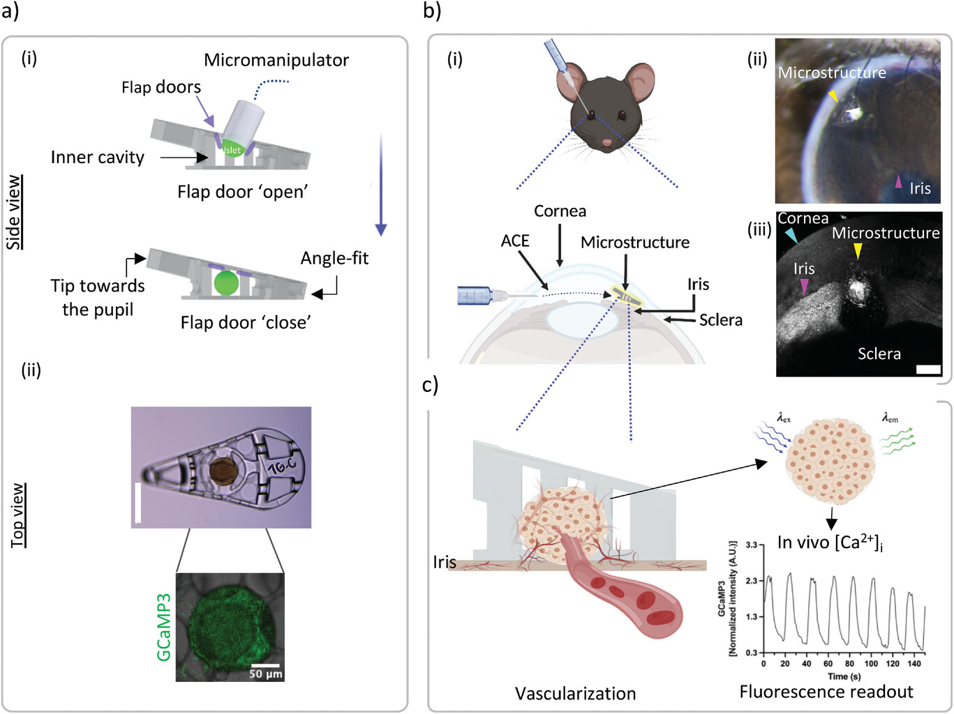
Wouter van der Wijngaart, a professor at the Department of Micro- and Nanosystems at KTH, explained the device’s unique design: “We have designed the medical device so that it can hold living mini-organs in a micro-cage with a new flap door technology, this to avoid the need for additional fixation.”
 Schematic overview of localization, transplantation, and application of pancreatic islet biohybrid microstructures. Image courtesy of KTH
This design means there’s no need for extra steps to keep it in place. Anna Herland, senior lecturer at the Department of Bionanotechnology at KTH, further highlighted: “We have designed the whole device as a wedge, and thus we can mechanically fix the structure in the angle between the iris and the cornea in the anterior chamber of the eye. When we tested the technology in mice, we observed that the device maintained its position in the living organism for several months and that the mini-organs quickly integrated with the blood vessels of the host animal and functioned normally.”
Harnessing 3D printing
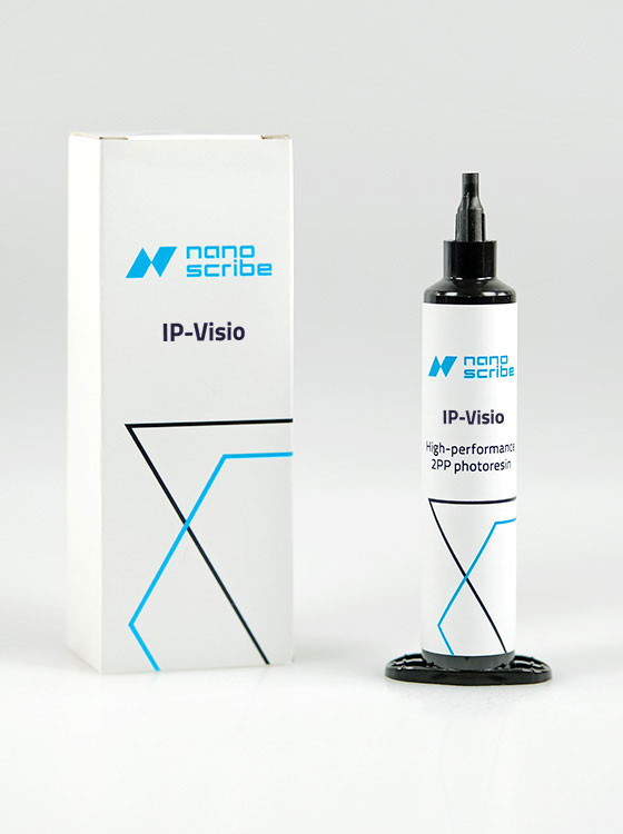
Central to this research is the 3D printed aspect of the microdevice. Titled 3D-Printed Biohybrid Microstructures Enable Transplantation and Vascularization of Microtissues in the Anterior Chamber of the Eye, the study emphasizes the power of merging biological components with precisely crafted structures. Nanoscribe (now under the BICO umbrella) developed the high-precision IP-Visio photoresin specifically for life sciences applications, and they use it to print these biohybrid microstructures.
IP-Visio is a special material made for 3D printing. It is exact, which is crucial for many scientific experiments. Plus, it’s safe for cells (non-cytotoxic) and doesn’t give off much glow, making it perfect for looking at cells under a microscope. Furthermore, it’s designed to work well with living tissues, which is crucial for the study’s goals.
Using Nanoscribe’s commercial two-photon polymerization (TPP) 3D printer, researchers tailor the microstructures to securely fit into the anterior chamber of the eye (ACE). This location is between the iris and the cornea. These structures ensure stability, even with the movement of the iris. With such a design, the device remains stable. During tests in mice, researchers transplanted tiny pancreatic sections into the ACE of a mouse’s eye.
 Nanoscribe IP-Visio photoresin. Image courtesy of Nanoscribe.
The mini-organs integrated with the host animal’s blood vessels, functioning normally for over 20 weeks. This approach not only highlights the usefulness of the device but also provides a deeper understanding of tissue interactions, vascular development in crafted tissues, and potential disease modeling. Notably, the researchers believe this is the first in vivo study of IP-Visio, emphasizing that comparisons with other transplantation studies are not possible.
Leap forward
The groundbreaking approach used in this research was under the expertise of Per-Olof Berggren, a professor of experimental endocrinology at Karolinska Institutet.
With vast experience in transplanting islets of Langerhans into the anterior chamber of mice eyes, Berggren stated, “The unit in question is unique and will form the basis for our continued work to develop an integrated microsystem for studies of the function and survival of the islets of Langerhans in the anterior chamber of the eye.” He also stressed the translational importance of this method, highlighting ongoing clinical trials involving human transplantation of islets of Langerhans in the ACE to treat diabetes.
Highlighting the broader importance of the research progress, Herland pointed out that cell transplantation treatments have begun in various disease areas, extending beyond diabetes. One of the main hurdles in improving cell treatments is not having a simple way to check how a transplant is doing. Without invasive procedures, it’s hard to guarantee the transplant will work in the long run.
“Our invention is a first step towards advanced medical microdevices that can both locate and monitor the function of cell transplants,” Herland states. “We have designed our device to position mini-organs, such as organoids and islets of Langerhans, without limiting the nutrient supply to the cells. Our work also demonstrates the first mechanical fixation of a device in the anterior chamber of the eye. Our design will enable future integration and use of more advanced device functions such as integrated electronics or drug release.”
Harnessing the capabilities of 3D printing, this research illuminates a path forward in medical science, especially when devising innovative strategies against challenges like diabetes. As the world confronts the escalating prevalence of diabetes, these biohybrid solutions could pave the way for a transformative approach to disease treatment and management.
Subscribe to Our Email Newsletter
Stay up-to-date on all the latest news from the 3D printing industry and receive information and offers from third party vendors.
Print Services
Upload your 3D Models and get them printed quickly and efficiently.


You May Also Like
Additive Manufacturing at a Crossroads
Additive manufacturing is at a crossroads. Simultaneously, we find ourselves between certain very different modalities, applications, and industries. Rather than being able to explore them all, companies will now have...
After 17 Years at 3D Systems, Katie Weimer Is Betting on Regenerative Breast Tissue
After spending 17 years helping build healthcare applications at 3D Systems and its predecessor Medical Modeling, Katie Weimer wasn’t planning to launch a startup. But when a regenerative breast tissue...
Why Elegoo Chose Emoji® to Introduce More People to 3D Printing
When Elegoo unveiled the world’s first officially licensed emoji®-themed 3D printer, it wasn’t just launching another version of an existing machine. The company was testing a much bigger idea by...
The Longevity Economy Needs a Factory
Longevity has become one of the biggest stories in healthcare. Every week seems to add a new announcement about an anti-aging therapy, an AI-powered drug discovery platform, or a startup...


