Cryogenic 3D Printing Improves Bioprinting for Bone Regeneration


Researchers from China continue in the quest to improve methods for bone regeneration, publishing their findings in “Cryogenic 3D printing of dual-delivery scaffolds for improved bone regeneration with enhanced vascularization.”
A wide range of projects have emerged regarding new techniques for bone regeneration—especially in the last five years as 3D printing has become more entrenched in the mainstream and bioprinting has continued to evolve. Bone regeneration is consistently challenging, and while bioprinting is still relatively new as a field, much impressive progress has been made due to experimentation with new materials, nanotubes, and innovative structures.
Cell viability is usually the biggest problem. Tissue engineering, while becoming much more successful these days, is still an extremely delicate process as cells must not only be grown but sustained in the lab too. For this reason, scientists are always working to improve structures like scaffolds, as they are responsible in most cases for supporting the cells being printed. In this study, the authors emphasize the need for both “excellent osteogenesis and vascularization” in bone regeneration.
In this case, the researchers employed cryogenic 3D printing, in which the phase change between liquid and solid is used to trigger polymerization to create extremely soft objects that can maintain their shape. The authors 3D printed dual-delivery scaffolds using a combination of β-tricalcium phosphate (β-TCP) and an osteogenic peptide (OP) containing composite emulsion inks. Cryogenic 3D printing has generally involved 3D printing a gelatinous material at a low temperature, such that it is deposited as a solid, or freezing the printed semi-liquid structure upon fabrication. These methods often result in more viable structures for tissue engineering and have become more popular in research over the past few years.
Here, the researchers agitated the TCP material for 30 minutes in an ice water bath (hence “cryogenic”) before combining it with the OP and printing it in a specialty printer. The resulting scaffolds were then freeze dried to remove any water and the composite emulsion inks with which the OP was mixed. The scaffolds were then coated in angiogenic peptide (AP) containing collagen, gelled for 30 minutes at 37 °C, and dried at room temperature. Whereas the use of OP was meant to promote osteogenesis, or bone growth, AP was meant to promote angiogenesis, the formation of new blood vessels.

Schematic illustration of fabricating AP and OP delivered TCP/PLGA composite scaffolds via cryogenic 3D printing and subsequent hydrogel coating. The scaffold can be implanted in bone defects to induce improve bone regeneration with required vascularization.
During characterization of the materials, along with in vitro and in vivo studies, the researchers found that the dual-peptide delivery scaffolds provided “improved new bone formation” when tested on rats, as well as providing improved vascularization in the tissue.
In studying mechanical properties, the researchers found that the collagen I hydrogel exhibited low compressive strength and elastic modulus, while the T and TB scaffolds showed strength and elastic modulus much closer to that of human bone.
“It is also found that the further coating of collagen I hydrogel onto the bone tissue engineering scaffolds (i.e., TV and TVB scaffolds), only a slight reduction in the compressive strength and elastic modulus was observed, suggesting that cryogenic 3D printed bone tissue engineering scaffolds coated with a thin layer of collagen I hydrogel are mechanically suitable for repairing/regenerating defected bone tissue,” explained the researchers.
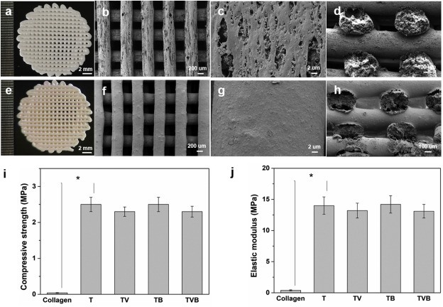
Morphology and mechanical properties of OP/TCP/PLGA and AP/collagen/OP/TCP/PLGA scaffolds. (a) and (e): digital images; (b-d, f-h): SEM micrographs of different scaffolds at different magnification; (i) compressive strengths of scaffolds and controls; (j) elastic modulus of scaffolds and controls. T, TV, TB, TVB refer to TCP/PLGA, AP/collagen/TCP/PLGA, OP/TCP/PLGA, AP/collagen/OP/TCP/PLGA, respectively.
The in vitro study showed a rapid AP release up to the 58 percent level for the TV and TVB groups, but it hit a plateau ten days later. OP being released from the TB scaffolds showed a “more sustained release level.” This was achieved at 79 percent after 42 days.

In vitro release behavior and degradation behavior of dual-peptide delivery scaffolds. (a) release behavior of angiogenic peptide and osteogenic peptide in a 42-day test period; (b) weight remaining of dual-peptide delivery scaffolds and controls in an 8-week test period.
In vivo studies showed that bone regeneration was only displayed in the peripheral area of the cranial defect in the rats. Defects with T and TV scaffolds displayed incomplete regeneration, while cavities within the TB scaffolds were filled with new bone tissue, aside from a gap in between scaffolds and the defects. TVB scaffolds showed “great improvement” with regeneration in the cranial defects.
Most likely, TVB scaffolds showed the greatest amount of regeneration due to the angiogenesis and osteogenesis brought on by the “synergistic effect” of AP and OP. Overall, in vitro angiogenesis was improved, while in vitro osteogenic differentiation was also enhanced.

Histological analysis of regenerated tissues in the rat cranial defects 3 months after the implantation of different scaffolds through Masson’s trichrome staining. Notes: “S” represents scaffold; “red arrow” indicates the vessel; “yellow star” indicates the new bone and osteoid. (For interpretation of the references to color in this figure legend, the reader is referred to the Web version of this article.)
Subscribe to Our Email Newsletter
Stay up-to-date on all the latest news from the 3D printing industry and receive information and offers from third party vendors.
Print Services
Upload your 3D Models and get them printed quickly and efficiently.


You May Also Like
PanOptimization Gets Attaboy from AFRL for PanX Software
Published in the International Journal of Advanced Manufacturing Technology, a paper titled “Part scale prediction of residual stress through thermomechanical modeling of additively manufactured Ti-6Al-4V” seems to point to the...
UCLA 3D Prints Zinc-Ion Battery With Seven Times More Energy
Just days after researchers at the California Institute of Technology unveiled a 3D printed design for lithium-ion batteries, another university team has announced a different battery breakthrough using additive manufacturing...
3D Printing News Briefs, July 1, 2026: Prosthetics, Drug Delivery, & More
We’re focused on healthcare and research in today’s 3D Printing News Briefs, including 3D printed prosthetics, patient-specific implants, drug delivery, and more. Read on for all the details! Students from...
New Study Shows Electronics Could Be Manufactured Directly in Space
A team of researchers from Auburn University and NASA Marshall Space Flight Center has successfully demonstrated a new additive manufacturing (AM) process that could allow astronauts to manufacture electronic components...