Chinese Researchers Sustain Bioprinted Brain Cells for Four Weeks

In the recently published ‘Engineering of brain-like tissue constructs via 3D Cell-printing technology,’ researchers use 3D models to glean information about neurology on numerous levels, from learning more about neural cells and monitoring the health of patients, to improving drug delivery.
As medical scientists and medical professionals—from clinicians to surgeons working on the cutting edge in terms of rare procedures—continue to use and improve anatomical models, patients benefit in many positive ways. A wide range of studies can be performed, doctors can make improved diagnoses and offer comprehensive treatments, medical students can practice without waiting for cadavers or other materials that may not always be highly accessible, and surgeons are able to plan surgeries better, along with using an array of new guides and tools.
Now, scientists from China are exploring bioprinting for neuroscience, and in vitro tissue constructs. While the benefits of cell culturing with the limits of 2D allow for a decrease in animal studies, taking it to the next level with 3D allows for much more substantial progress in research—especially due to greater sustainability of cells, which is one of the greatest challenges in studying and engineering of tissue.
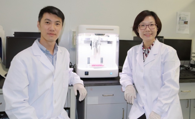
Yu Song (left) and Ting Zhang (right) from Tsinghua University are using 3D cell printing to engineer brain-like tissue constructs. (Courtesy: Biomanufacturing and Rapid Forming Technology Key Laboratory of Beijing at Tsinghua University)
“3D cell printing as an emerging biofabrication technology has been widely used to mimic natural 3D models in in vitro tissue research, especially in vitro brain-like tissue constructs in neuroscience,” state the researchers in their paper. “Fabricated brain-like tissue constructs provide a 3D microenvironment for primary neural cells to grow within. After more than several weeks of in vitro culturing, the formation of neural circuits in structures equips them with the capability of sensitively responding to a stimulus.”
With nutrients lying at the core of tissue viability, the researchers found success in this study while keeping cells alive for an impressive four weeks. Using a bioprinting ink that was rich in cells, they were able to deposit layers on electrodes, with spaces in between acting as areas for nutrients to feed the cells. Meanwhile, cell signals were monitored by the electrode arrays.
With their models serving as improved sources for drug testing and studying of neural networks, the scientists also realized survival of over three-quarters of the cells in the 3D structure. Such results lay in stark comparison to the 2D structure, displaying lack of survival for less than 25 percent of cells at the end.
The 3D model was created at Tsinghua University, at the Biomanufacturing and Rapid Forming Technology Key Laboratory of Beijing. Cells were suspended in gelatin, alginate, and fibrinogen—with 85 percent of them found to be sustained throughout the printing process. While emphasizing the need for proper nozzle diameter and print flow rate, the researchers were ultimately able to make bioink with mechanical properties so strong that they could actually mimic brain tissue.
“Our ongoing work includes studying how neuro drugs with different molecular structures diffuse at different speeds in our printed models, and administering electrophysiological stimulation and/or pentylenetetrazole to our models to study epilepsy,” said Wei Sun, director of the Biomanufacturing Center at Tsinghua University. “We are also interested in printing stem cells to build a brain-like model to study neurodevelopment and integrating the printed brain-like models with microfluidics to build a ‘brain-on-a-chip’ device.”
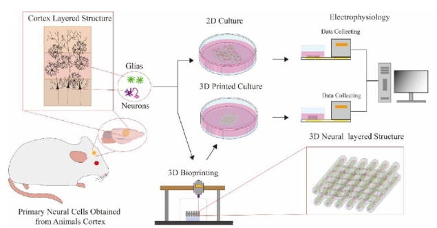
In 3D models, primary cells (collected from rats) are mixed with biocompatible materials to form bioink, and then printed on a petri dish for imaging or a 4×4 electrode array for electrophysiological recording. 2D samples are used as controls. (Courtesy: Biofabrication 10.1088/1758-5090/ab7d76)
What do you think of this news? Let us know your thoughts! Join the discussion of this and other 3D printing topics at 3DPrintBoard.com.
[Source / Images: ‘PhysicsWorld; ‘Engineering of brain-like tissue constructs via 3D Cell-printing technology’]Subscribe to Our Email Newsletter
Stay up-to-date on all the latest news from the 3D printing industry and receive information and offers from third party vendors.
Print Services
Upload your 3D Models and get them printed quickly and efficiently.


You May Also Like
The Stories nScrypt Can’t Tell; and Why That Matters
This article is Part 3 of a three-part series based on 3DPrint.com’s visit to nScrypt’s Orlando headquarters and conversations with Ken Church. There’s an interesting dynamic inside nScrypt’s Orlando headquarters. The...
amsight & toolcraft Improve AM Quality Control for the Semicap Market
As it is in the habit of doing at least once per generation, the semiconductor capital equipment (semicap) market is currently in the process of reinventing itself. This is too...
3D Printing Financials: XTPL Adds New Semiconductor and Defense Customers in Q1 2026
Polish microprinting company XTPL (WSE: XTP) reported first-quarter 2026 revenue of PLN 1.6 million (roughly $441,000) as the company expands into the semiconductor and advanced electronics markets, while also launching...
Creality Marks 12 Years with KliTek and AI-Powered Ecosystem Expansion
For 12 years, Creality has advanced accessible 3D printing technologies, enabling global users to turn ideas into tangible creations. What began as a desktop 3D printer manufacturer has evolved into...