
Using Symmetry and 3D Printed Medical Models to Repair Bone Fractures


In a new study, a team of researchers from China compared the clinical outcomes of treating isolated acetabular (concave surface of the pelvis) fractures with traditional 3D-printed planning models and using an approach that mirrors the affected area following the bilaterally symmetric nature of human anatomy. Their hypothesis is that, while 3D-printed models can definitely help when fixing hip fractures, mirroring hip bone models to gain a more accurate reconstruction of the pre-damaged bone might be more effective in its ultimate repair. This is important news in China, as the amount of “acetabular fractures and associated mortality” has been on the rise there for several years.
“Our findings are consistent with previous reports 1,5,7 and suggested that mirror model technology tends to improve the clinical outcomes for patients with acetabular fractures,” the researchers wrote.
They explained that for the most part, a human skeleton is “bilaterally symmetric; this is known as a mirror relationship.”
“This mirror relationship is the theoretical basis for using the contralateral acetabular mirror model as a template for the affected side. The mirror imaging model of the contralateral acetabulum replaces the post-reduction state of the affected acetabulum, which can greatly simplify the process of fracture assembly and reduction, improve the accuracy of reduction, and provide an excellent template for pre-bending of the pelvic reconstruction plate and determination of the screw length.”
Over the last few years, we’ve seen 3D printing grow more useful in the operating room and for custom fracture management. To complete a successful acetabular fracture surgery, strong internal fixation is necessary. The femoral head and acetabulum need to match, and the articular surface must have high integrity. 3D images are obviously better than CT data, but they’re still read on a 2D plane. This is why 3D-printed models can help.
“With the 3D model, we can accurately measure the degree of collapse of the articular surface, determine the number and shape of the fracture fragments, perform segmentation of each fragment model, separately print and assemble the fracture fragments, simulate reduction, and determine the position, length, and number of implants,” they stated.
The researchers used CT medical imaging data to print models of the contralateral acetabulum, then used the new 3D-printed mirror model “as a benchmark for surgical predrilling and determined the position and pre-bending properties of the pelvic reconstruction plate and the length of the screws.”
“We conducted a retrospective review of prospectively gathered data from our medical center from June 2011 to December 2017, in which consecutive patients who underwent traditional 3D printing technology or 3D printing mirror model technology following an isolated acetabular fracture were identified by the International Classification of Diseases (10th revision). Follow-up was initiated at the onset of the first day after primary acetabular fracture surgery,” they explained.
A total of 62 men and 84 women participated in the study. The patients treated with traditional 3D printing were in Group T, while those treated with 3D printing mirror model technology were in Group M.
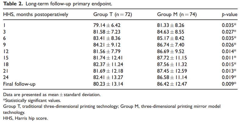
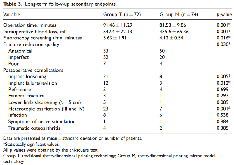
“In total, 146 advanced-age patients (146 hips) with an isolated acetabular fracture (Group T, n ¼ 72; Group M, n ¼ 74) were assessed for a mean follow-up period of 29 months (range, 24–34 months),” the team wrote. “The primary endpoint was the postoperative Harris hip score (HHS). The secondary endpoints were the operation time, intraoperative blood loss, fluoroscopy screening time, fracture reduction quality, and incidence of postoperative complications at the final follow-up.”
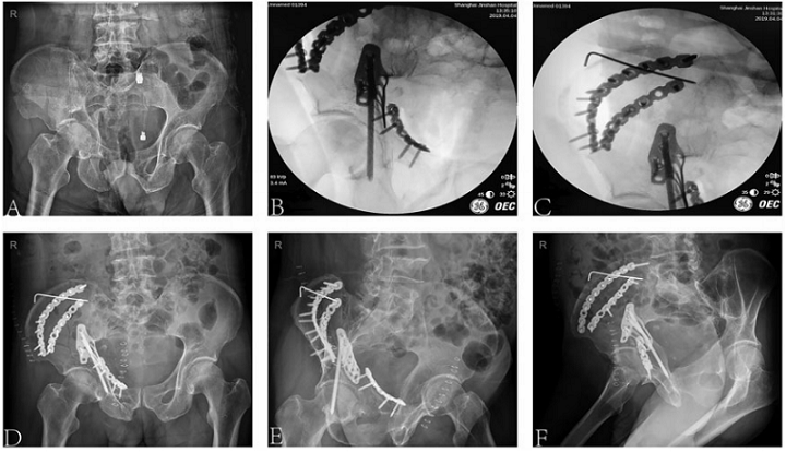
(a) Preoperative pelvic radiograph (anteroposterior view) taken at initial presentation revealing a right acetabular fracture (both columns). (b & c) Intraoperative fluoroscopic images of right acetabular fracture based on 3D printing mirror model technology. (d) Postoperative pelvic radiograph (anteroposterior view) and (e) anterior and (f) lateral images of the femur showing placement of the implants.
All patients needed certain criteria to be included:
- a unilateral isolated acetabular fracture
- fresh acetabular fracture with a 2-week duration from injury to operation
- no hip joint dysfunction or deformity pre-fracture
- able to understand instructions and follow rehab program
Materialise Mimics software was used for data processing, and the researchers directly generated 3D images of the affected acetabulum for Group T, while with Group M, “a 3D image of the affected acetabulum as well as a new 3D image of the contralateral acetabulum” were both generated. The STL files were sliced using Cura software and 3D printed on a Stratasys Objet 3D printer.
The same group of orthopedic surgeons operated on all of the patients.
“For Group M, we selected the appropriate reconstruction plate, pre-bent it to fit the bone surface, and adjusted it to the appropriate position; we then determined the specification and model of the pelvic reconstruction plate, the direction and length of the fixing screw, and the relationship of the acetabular joints,” they wrote.
The same intraoperative management was completed with the Group T patients, except before the reconstruction plate was chosen, the team removed the supports from between the bones in the 3D-printed model, and “dissociated each fragment to intuitively and accurately understand the fracture situation,” before resetting the fracture model.
“According to the preoperative plan, we completed the reduction as soon as possible, placed the pre-bent pelvic reconstruction plate in the position that had been established during the rehearsal, ensured that the plate and bone surface fit well, temporarily fixed the position, and inserted the appropriate screw into the predesigned screw trajectory on the 3D model to provide absolute stability of fracture reduction,” the team explained. “C-arm images were viewed in multiple planes to certify anatomic reduction.”
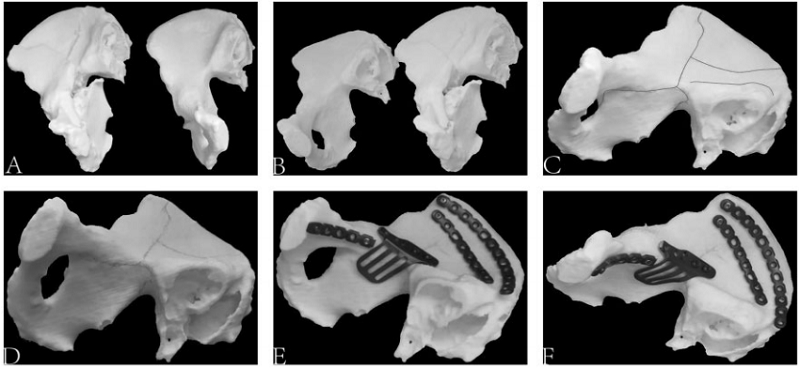
(a & b) 3D printed model of the bilateral acetabula providing an understanding of the normal 3D anatomy of the pelvic brim and acetabular columns. (c & d) Shape and orientation of the fracture line indicated by scribing on the mirror model. (e & f) 3D printed model with plate applied.
The patients all followed the same postoperative and rehabilitation instructions, such as wearing compression stockings for at least 21 days, completing continuous passive motion one day post-op, and touch-down weight-bearing with a walker between 2-30 days, and crutches between 30-90 days, post-op. Full weight-bearing was allowed after 90 days, based on clinical and radiography results.
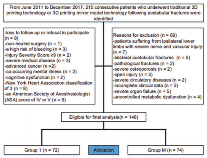
Study flow diagram.
“The patients were reviewed clinically and radiographically at 1, 3, 6, 9, and 12 months postoperatively and yearly thereafter,” the team stated. “The HHS, operation time, intraoperative blood loss, fluoroscopy screening time, and incidence of postoperative complications were significantly different between the groups, with Group M showing superior clinical outcomes.”
You can see the results of both the primary and secondary endpoints in the tables below.
“In conclusion, 3D printing mirror model technology in the treatment of acetabular fractures is associated with superior clinical outcomes compared with traditional 3D printing technology,” the researchers wrote. “The number of patients in this study was relatively limited, and it was not a prospective study; therefore, analysis of larger samples is needed for more accurate conclusions. Nevertheless, this study revealed accurate treatment of acetabular fractures through 3D printing mirror model technology, providing a new direction for the surgical treatment of acetabular fractures.”
Discuss this and other 3D printing topics at 3DPrintBoard.com or share your thoughts below.
Subscribe to Our Email Newsletter
Stay up-to-date on all the latest news from the 3D printing industry and receive information and offers from third party vendors.
Print Services
Upload your 3D Models and get them printed quickly and efficiently.


You May Also Like
How One Artist Is Using 3D Printing to Tell Stories About the Ocean
Artist Kimberly Callas sees something different when she looks at a 3D printer. Where others see a machine for making parts, she sees a way to tell stories about the...
Bambu Lab Wants Home 3D Printing to Feel Less Like a Workshop with PLA Pure
As desktop 3D printers become increasingly common in homes, Bambu Lab is focusing attention on something beyond print speed and hardware features. This week, the company launched a new filament,...
AM Asia Watch: China Exported 2.46 Million 3D Printers in Four Months
China’s consumer 3D printer industry seems to be reaching a new level of global dominance. According to Chinese state media outlet China Global Television Network (CGTN), China exported 2.46 million...
Bambu Launches A2L: What the New Printer Reveals About Its Strategy
Bambu Lab continues its relentless march for 3D printing domination with the launch of the A2L. The 330 × 320 × 325 mm printer will have a nozzle temperature of...


