
Chinese Randomized Trial Looks at How Useful 3D Printed Models Actually Are in Treating Bone Fractures


 Chinese researchers are examining how 3D printed models can improve treatment of fractures. Chunhui Chen, Leyi Cai, Wenhao Zheng, Jianshun Wang, Xiaoshan Guo, and Hua Chen of the Department of Orthopaedics at The Second Affiliated Hospital and Yuying Children’s Hospital of Wenzhou Medical University in Zhejiang explain their findings in ‘The efficacy of using 3D printing models in the treatment of fractures: a randomised clinical trial.’
We have followed cases previously where 3D printing has been used in creating prosthetics, fabricating arm casts to aid in mending broken bones, and making implants to heal injuries, but in this study the researchers evaluated the technology in terms of its ability to make improvements in pre-operative planning for breaks so serious that they require surgery. 48 patients suffering from distal radius fractures (one of the most common type of upper body injuries) participated in the study, with a 3D model made of each break.
The researchers then noted the following: operative time, amount of blood loss, and frequency of intraoperative fluoroscopy. Both surgeons and patients were given the opportunity to fill out questionnaires and offer input regarding the study at the end (among surgeons, the mean score for ‘usefulness of the 3D prototype for communication with patients’ was 9.1 ± 0.8 and that for ‘overall usefulness of 3D printing models’ was 6.7 ± 1.4.)
The scientists saw a need for this research project due to a high number of distal radius fractures and associated complications.
“Some authors have reported that the use of 3D printing models in the treatment of fractures has beneficial effects. Thus, we assumed that 3D printing models could be applied as a novel approach in the treatment of distal radius fractures,” states the research team. “As the anatomical volumes of distal radius are small, a realized model can be created conveniently, with little time and cost expenditure. In addition, the high incidence of such fractures enables the collection of data on standard cases. Thus, we believe that distal radius fracture is a suitable example with which to evaluate the efficacy of use of 3D printing models in the treatment of fractures.”
CT data was stored in DICOM format and then converted via Mimics software before 3D printing in PLA. The scientists reported success with the 3D printed models as they clearly displayed the fractures. Each surgery took around two and a half hours.
“On the 3D model, we split the fracture fragments according to the fracture line, and then temporarily reset the fragments with K-wire, followed by fixation of the fragments with metal plates and screws,” stated the research team. “In this way, the type and dimensions of the implant required were determined preoperatively, and we could choose suitable metal plates and screws.”
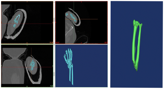
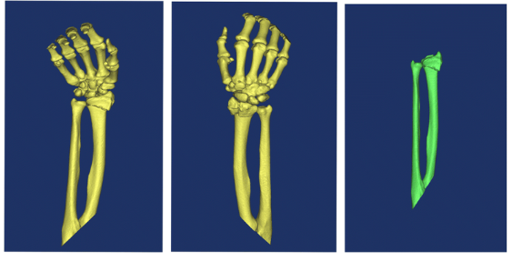
Reconstruction of a distal radius fracture in Mimics software v10.01
The researchers note in discussion that 3D printed models allow for more accurate diagnosis and surgery, along with serving as guides during procedures when needed.
“Because 3D printing can be used to produce an individualized, realized solid prototype of a fracture before complex surgery, junior surgeons can observe the anatomical structure of the fracture and simulate the surgical operation to determine the size of the implant required for internal fixation.”
The study also showed that 3D printed models allowed for faster procedure time, less blood loss in patients, and frequency of intraoperative fluoroscopy, but it ‘did not improve postoperative function.’ Limitations also include modeling of the bones only, without consideration to soft tissue, along with possible errors caused from the intricacies of software and hardware.
“Our study revealed that 3D printing models effectively help the doctors plan and perform the operation and provide more effective communication between doctors and patients, but cannot improve postoperative function compared with routine treatment,” concluded the researchers.
What do you think of this news? Let us know your thoughts! Join the discussion of this and other 3D printing topics at 3DPrintBoard.com.
[Source / Images: ‘The efficacy of using 3D printing models in the treatment of fractures: a randomised clinical trial’]
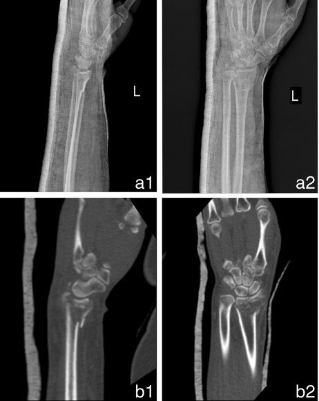
Preparation and outcomes of the surgical simulation. a1: The solid 3D model. a2: Preparation of the surgical simulation. b1, b2: Outcomes of the surgical simulation
Subscribe to Our Email Newsletter
Stay up-to-date on all the latest news from the 3D printing industry and receive information and offers from third party vendors.
Print Services
Upload your 3D Models and get them printed quickly and efficiently.


You May Also Like
Excellent Desktop Injection Molding, Made in Italy by Robot Factory
I was captivated when I saw my first Robot Factory 3D printer. The robust, precise machine was built to last. And this was in an era of very flimsy, disposable,...
Pogačar & Fairlight Cycles Show Us Low Cost 3D Printed Components for Bikes
There has been a lot going on in 3D printing for bicycles over the years. The most successful implementation so far is in bicycle seats. Carbon 3D printed seats are...
3D Printing News Briefs, June 18, 2026: Reseller, Relocation, Metal Space Powder, & More
We’ll start with business news in today’s 3D Printing News Briefs, as XJet appointed a value-added reseller in Germany, BIO INX is expanding its presence in the Italian market, and...
Researchers Combine AI and Bioprinting to Create Tiny Blood Vessel Networks
If 2026 has a theme in bioprinting, it may be blood vessels. Researchers can already print incredibly sophisticated tissues. The harder part is keeping those tissues alive. Without a network...


