
Shanghai Jiao Tong University School of Medicine: 3D Printing Scaffolds for Tendon-to-Bone Interface Engineering


Researchers from China’s Shanghai Jiao Tong University School of Medicine are exploring complex developments in tissue engineering, releasing their findings in the recently published ‘Three-dimensional printed multiphasic scaffolds with stratified cell-laden gelatin methacrylate hydrogels for biomimetic tendon-to-bone interface engineering.’
In fabricating a tendon-to-bone interface, the authors delve into a new method for improving biomechanics in the shoulder. Considering that rotator cuff tears are such a common issue—usually as a result of injuries, but also due to degenerative disorders—there is a need for improved techniques in restoring functionality to patients.
Currently, there are ‘significant challenges’ in surgical repairs; in fact, the researchers cite data that re-tears occur 20-90 percent of the time. With a biomimetic interface, there is the potential for overcoming current challenges—although previous attempts have yielded scaffolds lacking the proper optimization, along with the production of patches which have also not proved suitable for repair in more serious cases.
In this case, 3D printed scaffolds offer the benefit of:
- Customized structures
- Prevention of scaffold delamination
- Controllable pore sizes for better cell growth
- Use of poly(ε-caprolactone) (PCL), with excellent biocompatibility and biomechanical properties

(A) Illustration of the study design. (B) Photographs showing fabrication of the C/G-MS construct. FBs/GelMA was loaded on the PCL phase (a) and cross-linked under 405-nm visible light (b). Then, OBs/GelMA was loaded on the PCL/TCP phase and cross-linked (c and d). BMSCs/GelMA was injected into intermediate ducts (f) followed by cross-linking. Lateral views showing ducts before (e) and after injection (g). 3D = three-dimensional; BMSCs = bone marrow–derived mesenchymal stem cells; C/G-MS = cells/GelMA-multiphasic scaffold; FBs = fibroblasts; GelMA = gelatin methacrylate; OBs = osteoblasts; PCL = poly(ε-caprolactone); TCP = tricalcium phosphate; TGFβ3 = transforming growth factor β3.
To overcome previous challenges with cell seeding, the authors theorized that they could successfully seed cells of multiple types by using gelatin methacrylate (GelMA) hydrogels loaded on scaffolds in a stratified pattern. The scaffolds were developed to represent tendon, fibrocartilage, and bone. 3D printing was performed on a 3D-Bioplotter® Manufacturer Series printer.
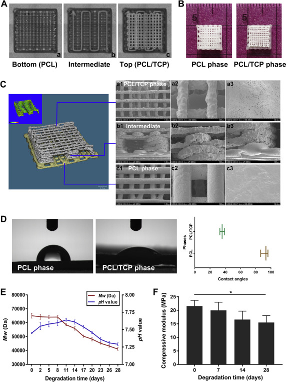
(A) Real-time images taken using 3D printing software during the bottom-up printing process, showing the 0/90°(a and c) and rectangular wavelike lay-down patterns (b). (B) Morphology and size of the 3D-printed scaffold. (C) MicroCT (left) demonstrated print quality and the predesigned structure. SEM micrographs (right) displaying surface microstructures of the PCL/TCP phase (a1∼a3), intermediate ducts (b1∼b3), and PCL phase (c1∼c3). (D) Contact angles separately obtained from PCL and PCL/TCP phases using the sessile drop method. (E) Changes in weight-average molecular weight (Mw) of the scaffolds and pH value of the incubation medium. (F) Compressive modulus of the scaffolds during 28 days of degradation. Data are mean ± SD. *p < 0.05. 3D = three-dimensional; microCT = microcomputed tomography; PCL = poly(ε-caprolactone); SD = standard deviation; SEM = scanning electron microscopy; TCP = tricalcium phosphate.
“A 0/90° lay-down pattern was used in the top (bone) and bottom (tendon) phases,” explained the researchers.
“This particular deposition pattern would eventually form four ducts with ample space for containing ECM-mimicking hydrogels and leave one opening of each duct for facile filling of hydrogels.”
Mice were used for the primary cultures in this study, featuring six males weighing 8 g each. Bone fragments were experimented with, including exposing the bone marrow cavity. After collecting cells, the researchers immersed the marrow with ‘growth media.’ These media were added further to promote cell attachment and growth, changed every three days, and treated with trypsin for passing.
Later in the study, the researchers attempted C/G-MS construct implantation by performing surgery on 27, 5-week-old mice.

Cytocompatibility tests. (A) Representative images of live/dead cell double staining of the C/G-MS construct on Day 1, 4, and 7. Scale bars: 100 μm. (B) Live/dead cell staining of both PCL and PCL/TCP phases exhibited good cell viability at Day 7. Scale bars: 500 μm. (C) Cell viability of both PCL and PCL/TCP phases was mostly over 90% at all time points. No significant difference was noted between the two phases. (D) The Cell Counting Kit-8 (CCK-8) assay showed an increase in cell proliferation in both C/G-MS constructs and cells/GelMA hydrogels during seven days in culture. Data are mean ± SD. C/G-MS = cells/GelMA-multiphasic scaffold; GelMA = gelatin methacrylate; PCL = poly(ε-caprolactone); SD = standard deviation; TCP = tricalcium phosphate.
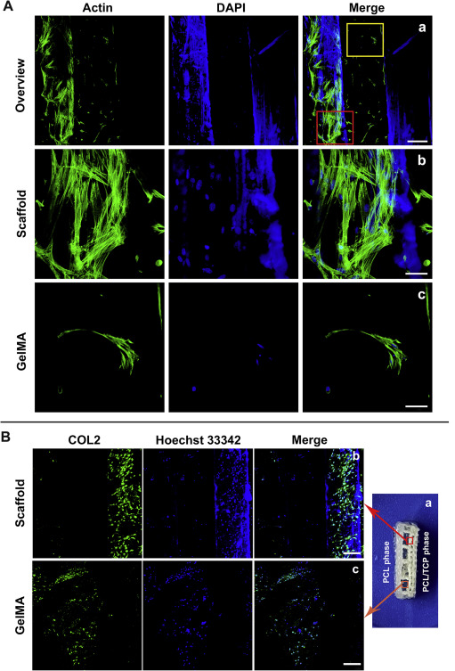
(A) Cellular actin staining in the C/G-MS constructs on Day 7. Red rectangle in (a) showing spreading of actin filaments in clustered cells on scaffold fibres (b), while yellow rectangle showing monolayered morphologies in GelMA (c). Scale bars: a, 200 μm; b and c, 50 μm. (B) Immunocytochemistry (ICC) analysis revealed chondrogenesis in C/G-MS constructs in vitro. COL2 emitting green fluorescence was detectable on the lateral of the intermediate phase (a), both on the surface of scaffold fibres (b) and in GelMA hydrogels (c). Scale bars: 200 μm. C/G-MS = cells/GelMA-multiphasic scaffold; GelMA = gelatin methacrylate; PCL = poly(ε-caprolactone); TCP = tricalcium phosphate.
“In our work, we managed to fabricate a biomimetic tendon-to-bone interface based on a much thinner 3D-printed multiphasic scaffold,” concluded the researchers. “The additive manufacturing technology not only provides rapid production via printing but also assures the integration and interconnectivity of the multiphasic scaffolds. The multihead 3D printing system used in this study allowed uninterrupted bottom-up printing from the biodegradable PCL phase mimicking the tendon to the osteoinductive PCL/TCP phase mimicking the bone.
“Our findings demonstrate that the stratified manner of fabrication based on the 3D-printed multiphasic scaffold is an effective strategy for tendon-to-bone interface engineering in terms of efficient cell seeding, chondrogenic potential, and distinct matrix deposition in varying phases,” concluded the researchers.
Materials for use in bioprinting and tissue engineering for scaffolds are the center of many studies that have the potential to change patients’ lives, from regenerating tissue after mastectomies to seeding human dermal fibroblasts, and experimenting with bone regeneration. What do you think of this news? Let us know your thoughts! Join the discussion of this and other 3D printing topics at 3DPrintBoard.com.
[Source / Images: ‘Three-dimensional printed multiphasic scaffolds with stratified cell-laden gelatin methacrylate hydrogels for biomimetic tendon-to-bone interface engineering’]
Subscribe to Our Email Newsletter
Stay up-to-date on all the latest news from the 3D printing industry and receive information and offers from third party vendors.
Print Services
Upload your 3D Models and get them printed quickly and efficiently.


You May Also Like
AM Asia Watch: Chinese Company Claims Advances in Titanium Powder Beyond 700C
They’re a familiar sight at trade shows: Chinese powder companies with barren stands lacking parts. There’s maybe some glass vessel with powder in it and a semi-complete data sheet, but...
Aires Tide Designed with AI, Supercomputers, and 3D Printing
The Department of Energy‘s National Nuclear Security Administration (DOE/NNSA) is part of the US government that manages the US nuclear stockpile, helping to upgrade, improve, and maintain nuclear weapons, and...
Largest Publicly Announced, Single Order in EOS History: Beehive Industries Spends $50M on M4 ONYX 3D Printers
Earlier this year, Beehive Industries received a $29.7 million contract to produce its Frenzy 6 and Frenzy 8 engines for the US Air Force. The metal additive manufacturing (AM) user...
Blue Origin’s New Glenn Explosion Comes During Major Manufacturing Push
Blue Origin‘s orbital New Glenn rocket exploded during a hot-fire test at Launch Complex 36 in Cape Canaveral on May 29, setting back the company’s launch ambitions at a time...


