

3D Printing Scaffolds for Regeneration of Tissue After Mastectomies & Tissue Damage


Researchers from Belgium and Germany explore topics in bioprinting, evaluating biocompatible structures in the recently published ‘Evaluation of 3D Printed Gelatin-Based Scaffolds with Varying Pore Size for MSC-Based Adipose Tissue Engineering.’ Focused on finding further progressive solutions for patients who have gone through mastectomies or other types of trauma involving soft tissue, the research team 3D printed and evaluated gelatin-based scaffold samples.
Because breast cancer is, unfortunately, both so common—and potentially devastating—to women around the globe, continued research is being performed regarding diagnosis, treatment, and procedures for the regeneration of soft tissue. Many of today’s methods, however, present obstacles—from microsurgical complications to high resorption rates. With the advent of adipose tissue engineering, there is the potential for regeneration through the integration of mesenchymal stromal cells (MSCs) and biomaterials.
In this study, photo-cross-linkable methacrylated gelatin (Gel-MA) is the material of choice, due to advantages such as its ability to interact with cells and similarities to collagen found in the extracellular matrix. The authors examine adipogenic differentiation behavior of bone-marrow-derived MSCs within 3D scaffolds that are porous. Rather than ‘confining’ cells, they allow three key activities: spatial spreading, movement, and distribution. With 3D printed samples, the research team was able to control parameters and make easy changes to 3D files and the resulting 3D prints.
Using an extrusion-based form of 3D printing, the team varied the sample scaffolds with strut spacing from 400 to 800 µm. This resulted in corresponding pore sizes of 230 ± 24 µm (400), 302 ± 30 µm (500), 348 ± 28 µm (600), and 531 ± 33 µm (800).

Physico-chemical characterization of the 3D printed Gel-MA scaffolds. a,b) Representative images of the pores together with the pore sizes obtained for the different scaffolds. The scale bars represent 200 µm. c,d) Stress versus strain curves obtained via compression tests from which the compressive modulus is determined. e) Mass swelling ratio of the scaffolds.
Control was maintained over high-shape fidelity scaffolds during printing by maintaining:
- Constant pressure (120 kPa)
- Temperature (30 °C)
- Writing speed (10 mm s−1)
The researchers also maintained consistency in UV exposure time and employed a high-precision nozzle (150 µm) to create stable scaffolds—all displaying similar strut widths. While all samples could absorb water, this ability was increased in those with larger pores. Overall, the team noted a connection between both swelling and mechanical properties of the sample scaffolds, due to the mass swelling ratio, which was increasing, and the compressive modulus which was decreasing.

Adipogenic differentiation of MSCs on extrusion-printed scaffolds. a) Gene expression of the adipogenic markers PPAR-γ, LPL, FABP, and FASN by MSCs on TCP compared to the scaffolds. b) Brightfield and c) immunofluorescent images showing clearly visible lipid droplets after 8 days of culture in adipogenic media. d) Quantification of adipogenic differentiation by normalizing Nile Red (stains lipid droplets) area to the number of nuclei. The scale bars represent 200 µm in all images except TCP panel in (c) where it is 100 µm.
“Although the stiffness of the extruded Gel-MA across all groups is 3–4 kPa mimicking native soft tissue compliance, the change in compressive moduli as pore size increases reflects the macroscale structural integrity of the scaffolds,” concluded the researchers. “In future work, maintaining low stiffness to promote adipogenic differentiation while improving the structural stability to improve implantation handleability could involve reinforcing the ink with secondary particles or phases.
“We found that MSCs differentiated robustly into the adipogenic lineage equally well in scaffolds of all pore sizes (200–600 µm). However, spatial distribution and cellular infiltration varied such that scaffolds with bigger pore sizes (>500 µm) support simultaneous differentiation and infiltration. These findings show the crucial importance of considering design parameters such as pore size when designing scaffolds for 3D soft tissue regeneration.”
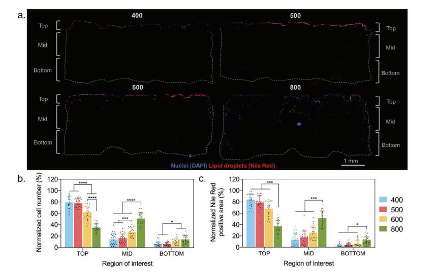
Spatial distribution of cells and lipid droplets in scaffolds. a) Representative scaffold cross sections stained with DAPI (nuclei) and Nile Red (lipid droplets) on each scaffold type showing differences in cell infiltration and spatial distribution of adipogenically differentiated cells. Quantification of b) cell and c) Nile Red positive area in different regions of the scaffold.
The study of bioprinting and scaffolding continues to expand, and while such research for diseases like breast cancer is critical, other scientists have published studies regarding uses for seeding dermal fibroblasts, promoting cartilage growth, 3D printing for bone replacement, and a variety of other ongoing projects within this very important field. What do you think of this news? Let us know your thoughts! Join the discussion of this and other 3D printing topics at 3DPrintBoard.com.
[Source / Images: ‘Evaluation of 3D Printed Gelatin-Based Scaffolds with Varying Pore Size for MSC-Based Adipose Tissue Engineering’]Subscribe to Our Email Newsletter
Stay up-to-date on all the latest news from the 3D printing industry and receive information and offers from third party vendors.
Print Services
Upload your 3D Models and get them printed quickly and efficiently.


You May Also Like
3D Printed Chip Packaging Specialist XTPL Enters Japanese Market
The additive manufacturing (AM) industry naturally wants to move beyond prototyping to production at scale, and the industry is certainly starting to demonstrate success with that objective, especially in Asia....
UT Researchers Use 3D Printing to Develop “Tabletop EUV Lithography” Process
Photolithography, the semiconductor manufacturing process whereby lasers transfer patterns onto chemical layers coating a substrate, is one of the most amazing industrial processes humanity has ever created. It is also...
3D Printing News Briefs, May 30, 2026: RIMPAC 2026, Acquisition, Ceramic Implants, & More
We’re kicking things off with materials news in this weekend’s 3D Printing News Briefs. Then it’s on to a hybrid manufacturing system for a maritime exercise, an expansion of industrial...
The University of Utrecht: “3D Printing Could Change Who Gets to Become a Manufacturing Power”
For decades, manufacturing has mostly been controlled by countries with huge factories, lower labor costs, and industrial systems that took years, sometimes decades, to build. But Utrecht University human geographers...


