Korea: Improving Implants for Knee Arthroplasty with Titanium Porous Coating in Direct Energy Deposition


Korean researchers are looking for ways to improve the materials used in total knee arthroplasty procedures. Design and technique have improved considerably in the past 30 years, but here the authors investigate the use of new materials for implants, outlining their findings in the recently published ‘Titanium Porous Coating Using 3D Direct Energy Deposition (DED) Printing for Cementless TKA Implants: Does it Induce Chronic Inflammation?’
While cementless fixation was meant to be a progressive technique promoting better bone to implant and growth and longevity, there have been concerns due to ‘poor clinical outcomes,’ as well as failure overall.
The researchers point out that there have been developments in 3D printing via direct energy deposition (DED) with the use of titanium. It may be considered ‘inferior’ to powder-bed-fusion; however, not for use in creating knee implants where it offers high mechanical strengths. In this study, the authors assess the use of Ti-coated on CoCr alloy, and how effective it is in stimulating an inflammatory reaction in both in vitro and in vivo models.
The team coated a CoCr substrate with a porous layer of pure Ti, creating a structure meant to match cancellous bone. Three samples were created, representing smooth and sand-blasted for comparisons and then DED surfaces as potential implants.
“Three types of specimens (n = 54; diameter: 14.6 mm; height: 3 mm fitted for 24 well plate), namely smooth (n = 18), sand-blasted (n = 18), and DED Ti-coated (n = 18), were manufactured for in vitro studies. Similarly, for in vivo studies, three types of specimen discs (diameter: 6 mm; thickness: 3 mm) were manufactured (n = 36): (1) smooth (n = 12), (2) sand-blasted (n = 12), and (3) DED Ti-coated (n = 12),” explained the researchers.

(A) schematic of a specimen used for experiments (A) smooth, (B) sand-blasted, (C) direct energy deposition (DED)-Ti coated (black color: cobalt chrome alloy, red color: Ti powder coating).
Thirty-six mice were added to the research project too, with each of them receiving the various sample implants.

(A) The experimental specimen was inserted into the dorsal subcutaneous layer. (B) After inserting the specimen into the mouse. Specimens were not inserted in the sham group after incision as the control group.
Viability of cells was examined on days 1,3,5, and 7, showing cytocompatibility for all surfaces. DED samples showed slightly elevated cell viability but not significant enough to make a difference. The researchers did note the same type of growth in ongoing experiments over the five-day period. There were no ‘statistically significant differences’ for in vivo immune profiling or in the concentration of inflammatory cytokines when compared to the sham group.

CCK-8 assay results for each specimen in a time dependent manner. The positive control, sand-blasted, and DED Ti-coated groups reached near their maximum value on day 7. However, the smooth group showed relatively low viability, but were not statistically significant (day 5 P = 0.1, Day 7 P = 0.1).
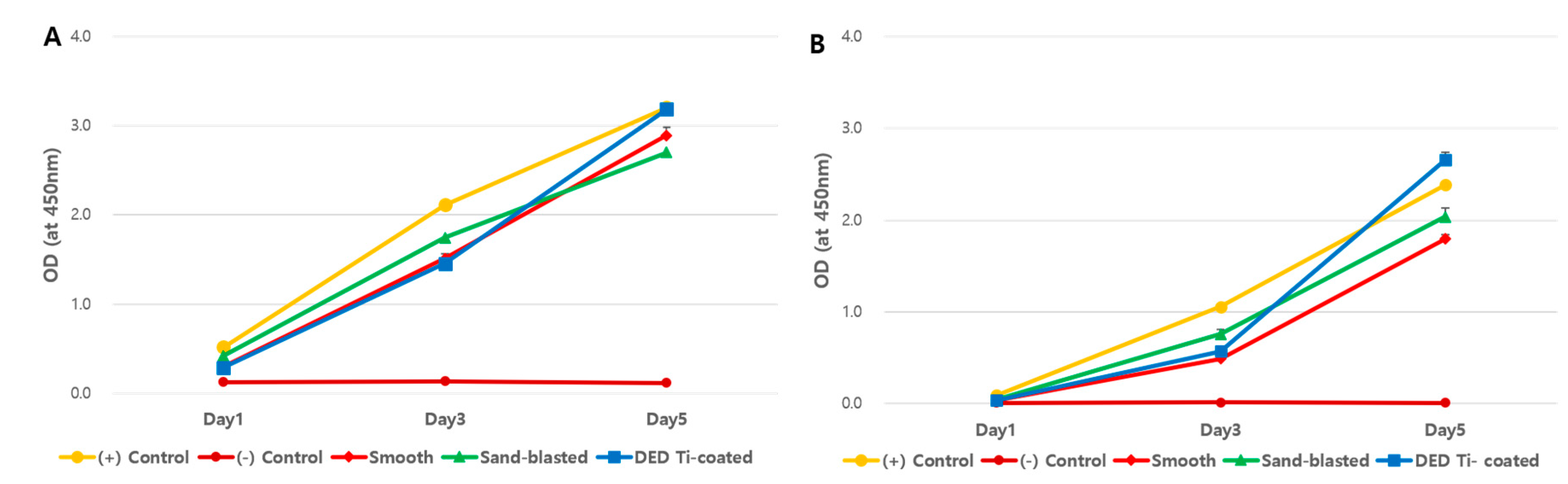
Results of re-testing using low ((A): 3 × 10³ cells) and high ((B): 1.2 × 10⁴ cells) concentrations of cells to determine the effects of initial cell concentration. All four experimental groups showed similar viability pattern and there were no statistically significant differences.
Overall, the researchers considered this process to offer great benefits, allowing it to surpass previous challenges in surface coating methods—with little chance for formation of Ti nanoparticles. Mechanical stability is high, and there is improved bonding between the Ti and CoCr alloys. Surface characteristics are easily manipulated for pore size, porosity, and the ultimate level of roughness. The researchers noted that DED techniques used in creating implants also show strong potential for decreasing ‘risk of inflammatory pathway.’
“… further research is needed to compare DED Ti-coated with TPS and PBF, which are the current technology used in vivo model. Finally, the inflammatory reaction in human and mouse could be different, thus there are limitations in applying our results obtained from mouse model directly to humans,” concluded the researchers.
“Moreover, our sample size was relatively small because of experimental ethics. In fact, most of the animal studies share these limitations, therefore our DED implant needs to be carefully evaluated on middle- and large-sized animal studies before clinical trial.”
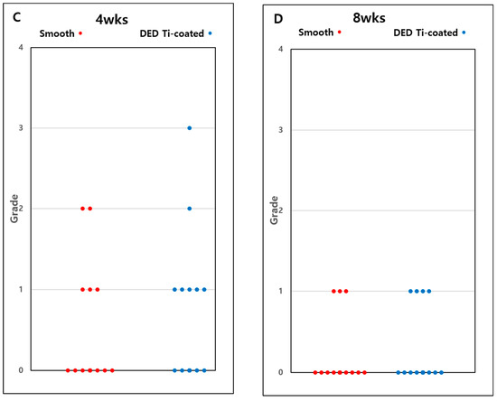
Evaluation of the degree of inflammatory cells by semi-Quantitative Grade System. Representative images of smooth (A) and DED Ti-coated (B) at 4 weeks after implantation. Macrophage (Green arrow head), lymphocyte (blue arrow), granulocytes (white arrow heads), and monocytes (white arrow) were observed in both specimens, however, not more than 10% of all cells. Both representing zone evaluated as grade 1. The result of semi-Quantitative Grade analysis represented at 4 weeks (C) and 8 weeks (D). The expression level of inflammatory cells was decreased at 8 weeks (D) compared with 4 weeks (C), however, there was no statistically significant difference (smooth group P = 0.413, DED Ti-coated group P = 0.219). There was no significant difference between the smooth and DED Ti-coated groups at 4 (P = 0.551) and 8 weeks (P = 0.755).
3D printed implants and other devices are used for a wide range of procedures today, from the fabrication of 3D printed medical models for hip surgeries, to pre-planning systems for shoulder surgeries, total knee replacements, and more.
What do you think of this news? Let us know your thoughts! Join the discussion of this and other 3D printing topics at 3DPrintBoard.com.
[Source / Images: ‘Titanium Porous Coating Using 3D Direct Energy Deposition (DED) Printing for Cementless TKA Implants: Does it Induce Chronic Inflammation?’]Subscribe to Our Email Newsletter
Stay up-to-date on all the latest news from the 3D printing industry and receive information and offers from third party vendors.
Print Services
Upload your 3D Models and get them printed quickly and efficiently.


You May Also Like
Goal! 3D Printing for the 2026 FIFA World Cup
The 2026 FIFA World Cup officially kicked off in Mexico City on June 11th. It’s the largest FIFA tournament in history, with 48 teams competing over 104 matches. Instead of...
Bambu Lab Wants Home 3D Printing to Feel Less Like a Workshop with PLA Pure
As desktop 3D printers become increasingly common in homes, Bambu Lab is focusing attention on something beyond print speed and hardware features. This week, the company launched a new filament,...
The Rise of IP: The First Emoji 3D Printer Is Here. Don’t Rule Out Star Wars Next.
For years, most desktop 3D printers have looked more or less the same. Some are black. Some are gray. A few are bright orange. They look like boxes. Some are...
AM Asia Watch: China Exported 2.46 Million 3D Printers in Four Months
China’s consumer 3D printer industry seems to be reaching a new level of global dominance. According to Chinese state media outlet China Global Television Network (CGTN), China exported 2.46 million...