3D Printing Microneedles for Biomedical Applications

In the recently published ‘Biocompatible 3D Printed Microneedles for Transdermal, Intradermal, and Percutaneous Applications,’ authors Khalil Moussi, Abdullah Bukhamsin, Tania Hidalgo, and Jurgen Kosel explore the use of microneedles in many different medical applications—mainly for procedures that are considered only minimally invasive.
Microneedles are being used today in drug delivery as they can assist in diagnosing and delivering drugs to necessary areas. Numerous materials have been used to date—to include glass, silicon, glass, plastics, and metals. Numerous techniques have been attempted also. The authors point out that they have in some cases even been used in ‘complex microsystems,’ although in general they are used as devices that stand alone.
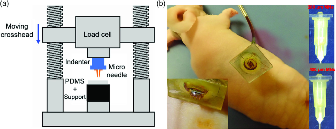
MN penetration testing. a) A schematic view of the setup used for PDMS skin‐like samples. b) In vitro skin piercing experiment.
“Despite their perceived advantages and applications, MNs are yet to see an extensive pervasion in clinical settings. In part, this is due to the complexities associated with the microfabrication techniques used for the development of MNs, which are often multistep, labor intensive, and require expensive cleanroom equipment,” explain the researchers. “In addition, complications arise from the handling and attachment of MNs fabricated with a specific process to a device or structure fabricated with another process.”
3D printing continues to become more of an option in creating MNs, especially through greater evolution of and introduction of SLA and FDM 3D printing and two‐photon polymerization (TPP). FDM 3D printing has been more commonly used so far, and especially with recent projects such as fabrication of biodegradable polymer MNs for transdermal drug delivery. TPP shows potential, however, for 3D printing hollow MNs meant for transdermal delivery in patients, and for this study, the researchers focus on how to attach such MNs to a drug reservoir.
“This approach has already been implemented using standard processing techniques of silicon microfabrication and using metal electrodeposition of a patterned sacrificial mold to create hollow MNs with an attached reservoir,” state the researchers. “However, these processing techniques suffer from the same complexities as mentioned earlier and afford limited control over the MNs geometry and aspect ratio. Thus, with the advent of TPP, it is now feasible to 3D print microstructures having dimensions in the millimeter scale while providing feature sizes with micrometer resolution.”
Overall, results showed ‘excellent linear fit’ as well as FEM simulation. The researchers recognized that the study shows the potential material strength necessary, along with the ability to tailor elasticity through changing parameters like slicing and hatching. The research team created a reservoir of 2 mm3 volume topped with hollow MNs with inner diameter and height ranging from 80 to 120 μm and from 200 to 400 μm.
“Combined with experimental testing, this analysis verified the appropriate dimensions of the MNs that are needed to ensure mechanical stability for a given application. To corroborate the applicability of the 3D printed MNs, they were used for a penetration test into both a skin‐like material and mouse skin. Penetration into skin‐like material allowed the determination of the piercing force which was 0.095–0.115 n,” stated the researchers.
“Confocal microscopy of the mouse skin confirmed the MN array penetration and fluorescent dye delivery 100 and 180 μm deep into the skin for the 200 and 400 μm‐long MNs, respectively. A complementary biocompatibility assessment was performed to investigate the potential of using the technique for direct tissue interfacing or implants, and it has determined that the photoresist has minimal cytotoxicity, which makes it ideal for such applications.”
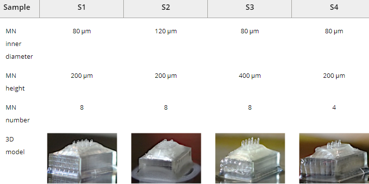
Dimensions of the 3D printed specimens used for the fluidic characterization
The study of microneedles continues for researchers, along with continued innovation such as MNs that dissolve under the skin, deliver drugs and communicate through BlueTooth, allow for new types of arrays. What do you think of this news? Let us know your thoughts; join the discussion of this and other 3D printing topics at 3DPrintBoard.com.
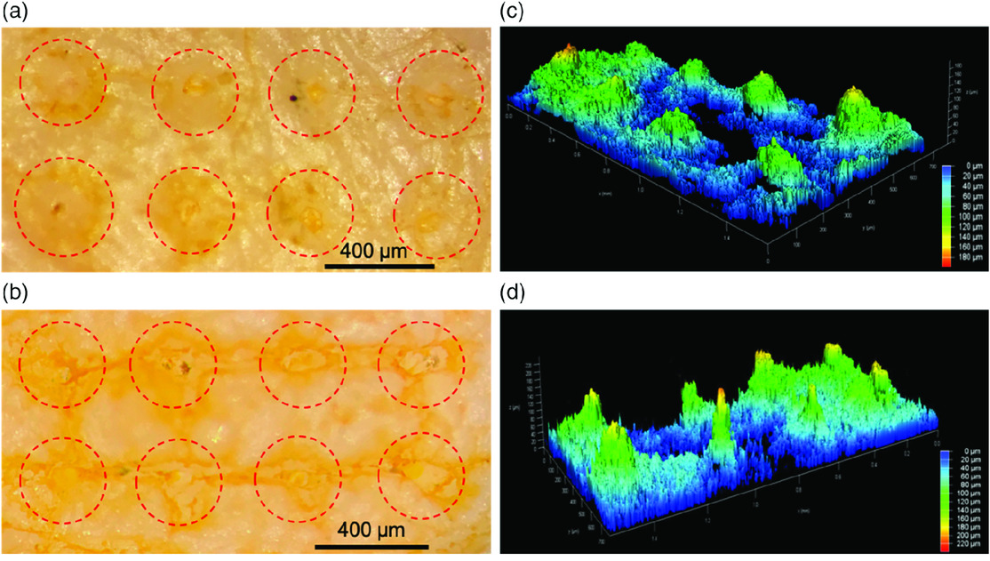
MNs penetration and delivery of FITC dye into mouse skin. a,b) Photograph of the skin after penetration and dye delivery using 200 and 400 μm‐long MN arrays, respectively. c,d) 3D representation of dye delivery in the mouse skin for the 200 and 400 μm‐long MN arrays, respectively. The blue side (z = 0) corresponds to the skin surface, and z > 0 represents the penetration into the skin.
Subscribe to Our Email Newsletter
Stay up-to-date on all the latest news from the 3D printing industry and receive information and offers from third party vendors.
Print Services
Upload your 3D Models and get them printed quickly and efficiently.


You May Also Like
3D Printing News Briefs, May 9, 2026: Financials, Large-Format Printer, Steels, & More
In 3D Printing News Briefs this weekend, 6K Additive has appointed a new COO and released its Q1 financials. Rolls-Royce opened a new AM Development Cell, and MODIX launched its...
LOOP 3D Reveals 3D Printer Designed for Industrial Uptime
LOOP 3D has released the LOOP PRO X+ TURBO Gen2. This version has been improved to meet the needs of manufacturing clients. The printer is meant to be a production unit...
3D Printing News Briefs, April 25, 2026: Competition Winners, AI Platform, X2D Printer, & More
In this weekend’s 3D Printing News Briefs, AMUG announced the winners of its Technical Competition, and Authentise launched AI platform Whisper at RAPID. Bambu Lab wasn’t at RAPID, but launched...
HP Continues to Lower Barriers to Adoption with Compact MJF 1200 & Other RAPID + TCT Announcements
This week at RAPID+TCT in Boston, HP Additive Manufacturing Solutions is celebrating ten years in the AM market. The company launched its Multi Jet Fusion 3D printing technology in Barcelona...