

Sichuan University Researchers Examine Four Levels of 3D Printed PCL Scaffolds


3D printing in medicine and in structures like scaffolding has become almost commonplace today, but scientists continue to refine the processes to help patients with a variety of conditions, many of which are life threatening. A team from the Research Center for Nano-Biomaterials at Sichuan University has recently decided to explore an important and relevant topic in ‘Modification of 3D printed PCL scaffolds by PVAc and HA to enhance cytocompatibility and osteogenesis.’
Four groups of scaffolds were 3D printed in the study, in the following materials:
- PCL
- PCL/PVAc
- PCL/HA
- PCL/PVAc/HA
The team studied their morphologies, mechanical properties, and biological characteristics, with two new types of bone formation patterns discovered during the study—one formed on the grid matrix and another as new bone remolded into circles after previously being formed in the middle of the structure.
In the introduction to their study, the researchers discuss the potential 3D printing offers for material scientists and orthopedic surgeons, as scaffolds can be made layer by layer in accordance with available and personalized CT data exhibiting bone defects.
“With the help of 3D printing technology, clinicians can fabricate scaffolds of different size, specific shape and porosity,” state the researchers.
PCL has been approved by the FDA and offers a suitable material for 3D printing scaffolds due to high crystallinity and a low melting point. The research team points out that it also offers ‘superior workability and machinability’ when printing at normal temperatures. The materials are already in use for a wide range of medical needs, including cranial repairs, screws to fix bone fractures, systems with sustained-release mechanisms, and already for use as a 3D printing matrix for hydroxyapatite scaffolds. PCL is also a bioabsorbable material that is deemed exceptionally safe.
With all the good of course, comes some ‘bad,’ and the team of scientists discusses the shortcomings they uncovered with PCL and hydroxyapatite (HA):
“On one hand, PCL is a hydrophobic material with no propensity for cell attachment. On the other hand, the degradation rate of PCL is very slow. A polymeric shell naturally forms on the stent of scaffolds during the printing process. Thus, it is difficult to expose the embedded HA particles of PCL matrix, which affects the bone-bonding function of HA material.”
The team goes on to point out the need for the following features in scaffolds:
- Controlled biodegradability
- Appropriate mechanical strength
- Interconnected pore structure
- Porosity for cell in-growth
3D printing is considered suitable for all these requirements because it allows for fabrication of the interconnected pores for bone regeneration.

(a) The implantation process of a 3D printed scaffold into bone defects of rabbits. (b) Microstructure of human cortical bone. (c) Schematic diagram of the channel structure which is an ideal space for bone tissue ingrowth; a channel structure has been observed along with the black arrow. (d) Gross photo of the 3d-printed scaffold (e) photo of the side of printed scaffold. (f) Photo of the top surface of printed scaffold. (g) the channel shown in the sketch map of the 3d-printed scaffold structure.
The four scaffold groups were 3D printed on the 3D Bioprinter V2.0 (manufactured by Hangzhou Regenovo Biotechnology Co., Ltd, China), all on the same settings:
“0.34 mm diameter nozzle, a filament gap of 0.5 mm, a layer thickness of 0.1 mm, and a lay-down pattern of 0°/90°. The size of a filament gap was set as 0.5 mm in agreement with previous studies.14 Disc scaffolds of 14(D) × 1.5(H) mm were fabricated for in vitro cell culture. Column scaffolds of 5(D) × 6(H) mm were fabricated for in vivo studies. Rectangular scaffolds of 10(L) × 10(W) × 10(H) mm were fabricated for mechanical tests and other characterization analyses.”
The results showed that all scaffolds had similar porosity in the range of 74.1 percent to 76.1 percent, but there were differences in mechanical properties:
“The PCL scaffold held the highest compressive strength of 11.9 MPa (p < 0.05), while the PCL/HA scaffold had the highest modulus of 125.4 MPa (p < 0.05). The PCL/PVAc scaffold showed the lowest values of both the compressive strength (3.9 MPa) and modulus (26.6 MPa). The values of mechanical properties of PCL/PVAc/HA tri-component scaffold (6.3 MPa and 55.5 MPa) were higher than those of PCL/PVAc but lower than those of PCL and PCL/HA with no statistical difference observed among them.”
Overall, the scaffolds were successful, although the PCL/PVAc/HA scaffold showed more favorable characteristics during in vitro cell culture experiment and in vivo bone formation.
“The new 3D printed scaffold presents a promising prospect for future biomedical applications,” concluded the researchers.

H&E stained histological section images of PCL/HA and PCL/PVAc/HA scaffolds after implantation in bone defects for 4, 8 and 12 weeks.
Find out more about this study here. And if you are interested in finding out more about 3D printed scaffoldings, follow some of our other stories on topics like thermoresponsive nanohybrid scaffolds, lattices made in 3D printed rectangle form, and even neural scaffolds.
What do you think of this news? Let us know your thoughts! Join the discussion of this and other 3D printing topics at 3DPrintBoard.com.
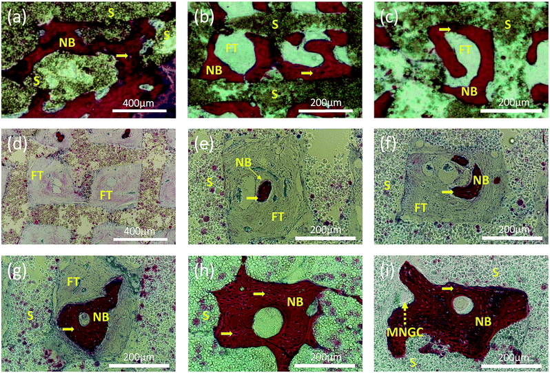
Representative histological section images stained by H&E showing two patterns of new bone formation: one pattern represents the new bone growing close to the wall of the grid (a–c); another pattern represents the new bone, emerging early in the center of the grid and then growing and filling the entire grid with a channel in the center (d–i), similar to the natural osteon formation. Pictures (a–c) are of the samples of PCL/HA scaffolds after 12 weeks implantation. Pictures were taken of the samples of PCL/PVAc/HA scaffolds after implantation for 4 (d and e), 8 (f and g) and 12 (h and i) weeks, respectively. Yellow arrows: osteocytes; yellow dashed arrow: multinucleated giant cell; NB: new bone; S: scaffolds; FT: fibrous tissue; MNGC: multinucleated giant cell.
Subscribe to Our Email Newsletter
Stay up-to-date on all the latest news from the 3D printing industry and receive information and offers from third party vendors.
Print Services
Upload your 3D Models and get them printed quickly and efficiently.


You May Also Like
3D Printing News Briefs, June 18, 2026: Reseller, Relocation, Metal Space Powder, & More
We’ll start with business news in today’s 3D Printing News Briefs, as XJet appointed a value-added reseller in Germany, BIO INX is expanding its presence in the Italian market, and...
Scientists Use BMF to 3D Print Seal Whiskers That Track Prey Long After It’s Gone
Seals use their whiskers to hunt. Not Navy Seals, although they may in some way also, but this article is about lowercase seals. Not Seal the musician either; as far...
Researchers Combine AI and Bioprinting to Create Tiny Blood Vessel Networks
If 2026 has a theme in bioprinting, it may be blood vessels. Researchers can already print incredibly sophisticated tissues. The harder part is keeping those tissues alive. Without a network...
University of Arkansas Researchers Test Metal 3D Printing in a Mars-Like Atmosphere
If humans eventually establish a long-term presence on Mars, they will face a major manufacturing challenge almost immediately. Tools will break. Parts will wear out. Equipment will need repairs. But...


