

Valentines Special! Researchers in India Explore 3D Printing Heart Valves


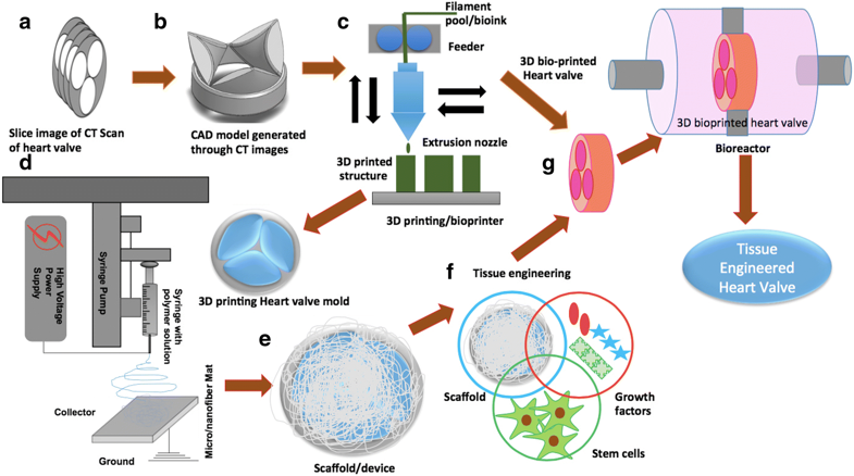
Schematic representation of the proposed process for the generation of 3D heart valves through combining either bioprinting or combination of 3D printing and electrospinning with bioreactor to arrive at functional tissue engineered heart valves (a) slice of CT images, (b)3D CAD model generation, (c) 3D bioprinting through bioink/ 3D printing through PLA, PCL materials, (d) combining PCL-Gel electrospun nanofibrous with 3D printed scaffold, (e) scaffold ready for conventional tissue engineering (f) Development of tissue through combining stem cells, growth factors and developed scaffold, (g) Development and initial tissue remodeling in perfusion bioreactor under dynamic pulsatile flow conditions
The heart is central to keeping you alive, pumping blood, oxygen, and nutrients throughout your body—and eliminating waste too. Disease of this central organ is also one of the leading causes of deaths in humans, with coronary heart disease most common. Hundreds of thousands of individuals die each year from heart-related issues, and over 700,000 individuals in the US have heart attacks.
Researchers continue to find better ways to prevent such disease, along with saving patients who are in peril after experiencing cardiac abnormalities. Enter 3D printing, and new research from India, as scientists Rajat Vashistha, Prasoon Kumar, Arun Kumar Dangi, Naveen Sharma, Deepak Chhabra, and Pratyoosh Shukla publish their findings in ‘Quest for cardiovascular interventions: precise modeling and 3D printing of heart valves.’
The authors are encouraged not only by the ‘digitalization of health care practices’ today but also by 3D simulation and computational modeling assisting in surgeries. With these progressive methods in place and becoming more frequently used by medical professionals around the world, the scientists see 3D printing also as having great potential for helping to resolve valvular problems—especially as bioprinting of tissues (and organs, eventually) continues.
The scientists list the following issues as the most common leading to valvular heart diseases (VHDs):
- Aortic regurgitation
- Aortic stenosis
- Primary mitral regurgitation
- Secondary mitral regurgitation
- Mitral stenosis
- Tricuspid regurgitation
- Tricuspid stenosis along with coronary artery disease,
- Rheumatic fever
- Bacterial endocarditis
“These VHDs are associated with significant morbidity and mortality in an aged population, as they are correlated with vascular disorders,” state the researchers. “Considering the reasonable percentage of aged population in Europe, North America, Japan and other countries, VHDs are one of the prominent causes of death in these regions and need immediate attention.”
The team states that prosthetic valve replacement, by means of either mechanical or biological valve, is the ‘only exclusive solution’ possible today. There are still problems with these types of valves, however, due to issues with leaking, the need for excessive care, medication, and continued imaging by specialists. Diagnosis can often be overly invasive too, and the researchers point out that improvements can be made with the use of 3D technology not only in diagnosing but also in simulations used in establishing alternative therapeutics.
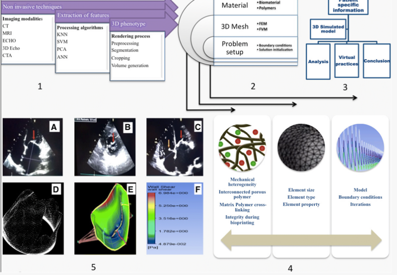
Schematic representation of the cardiovascular modeling process for patient specific diseases diagnostics. Processes 1, 2 and 3 show the sequential steps whereas step 4 and 5 shows conditions for real time processing. a. Thick and calcific Mitral valve with decreased opening in case of Chronic Rheumatic Heart Disease, (b). Parasternal Short Axis view of Mitral valve showing thickened anterior and posterior leaflets with reduced valve area, (c). Four Chamber view showing thickened Tricuspid Valve (yellow arrow) suggestive of organic Tricuspid valve disease and thick and calcific Mitral valve (red arrow) in case of Rheumatic Heart Disease, (d). 3D mesh for the volume generated geometry. e. Numerical setup for the problem in CFD software, F. Result post processing.)
Previous efforts at creating artificial heart valves have been rife with challenges, leaving the researchers to state:
“Henceforth advancements in imaging, computational modeling and designing tools need to be integrated with emerging areas of tissue engineering in order to develop human prosthesis similar to native tissues. Tissue engineering holds the potential to reduce patient–prosthesis mismatch in the direction of personalized medicine and accelerate the design and developmental time of prosthetic devices.”
The creation of a heart valve via tissue engineering allows the artificial material to mimic the ‘native valve’ as it is implanted. Also, a 3D printed model gives medical professionals the opportunity to understand tissue biology and more about how a patient’s disease is progressing—and what therapeutic interventions might be effective.
“Before tissue engineering a heart valve, it is imperative to understand the multi-scale architecture, geometry and biomechanics of a heart valve’s parts that play a significant role in remodeling of a neo tissue matrix in the dynamic mechanical environment of a functional heart valve,” state the researchers. “These understandings will enable proper selection of biomaterials, fabrication methodologies, characterization tools and developmental environments for generation of tissue engineered heart valves (TEHVs).”
3D printing via extrusion is not the best choice for creating heart valves, nor is ceramic based 3D printing or SLA; however, bioprinting and inkjet technology may be best suited, along with materials bordering on the 4D realm, inspired by origami. The key in the future will be to refine tissue engineering bioprinting further to ‘eradicate potential defects in prosthetic heart valves,” concluded the researchers.
What do you think of this news? Let us know your thoughts! Join the discussion of this and other 3D printing topics at 3DPrintBoard.com.
[Source / Images: Quest for cardiovascular interventions: precise modeling and 3D printing of heart valves]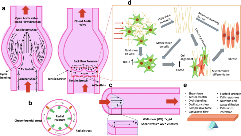
Schematic representation of the forces acting on the aortic valve during pulsatile blood flow and remodeling of fibrous matrix by cells of aortic valve under the influence of blood shear for open and closed state along with the factors responsible for the balanced state (a) representation of valve in frontal plane, (b) transverse cross-section view of blood vessel, (c) longitudinal cross-section view of the blood vessel, (d) fibrous matrix remodeling by cells and (e) balancing of factors while developing TEHVs
Subscribe to Our Email Newsletter
Stay up-to-date on all the latest news from the 3D printing industry and receive information and offers from third party vendors.
Print Services
Upload your 3D Models and get them printed quickly and efficiently.


You May Also Like
3D Printed Chip Packaging Specialist XTPL Enters Japanese Market
The additive manufacturing (AM) industry naturally wants to move beyond prototyping to production at scale, and the industry is certainly starting to demonstrate success with that objective, especially in Asia....
UT Researchers Use 3D Printing to Develop “Tabletop EUV Lithography” Process
Photolithography, the semiconductor manufacturing process whereby lasers transfer patterns onto chemical layers coating a substrate, is one of the most amazing industrial processes humanity has ever created. It is also...
3D Printing News Briefs, May 30, 2026: RIMPAC 2026, Acquisition, Ceramic Implants, & More
We’re kicking things off with materials news in this weekend’s 3D Printing News Briefs. Then it’s on to a hybrid manufacturing system for a maritime exercise, an expansion of industrial...
The University of Utrecht: “3D Printing Could Change Who Gets to Become a Manufacturing Power”
For decades, manufacturing has mostly been controlled by countries with huge factories, lower labor costs, and industrial systems that took years, sometimes decades, to build. But Utrecht University human geographers...


