Artec 3D Scanners Help with Creation of Customized Chest Implants

Based in France, AnatomikModeling designs and develops custom 3D printed implants, such as the world-first custom 3D printed airway stents it developed last year. One of the company’s main areas of focus is on congenital thoracic deformities, or anomalies in the growth of the chest wall. These deformities include pectus excavatum, which is an excessive growth of the cartilage that joins the ribs to the breastbone and causes a concave defect of the sternum, and Poland syndrome, which results in an underdeveloped chest muscle on one side of the body.
Conditions like these are prime targets for the kind of personalized, low invasive treatment that is AnatomikModeling’s specialty. Customized medical devices can more effectively treat complex anatomical defects than their traditional, one-size-fits-all counterparts. AnatomikModeling’s CTO and Co-Founder Benjamin Moreno and his team have been using Artec 3D scanning solutions and Geomagic Freeform software to design custom chest implants and monitor the results.
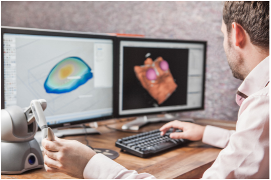
Before undergoing an operation, a patient will have several scans taken to gather the required data on the deformity. Artec 3D scanners capture the data of the outside of the chest, while CT scans reveal the internal structure of the rib cage. Data from the CT scan is then imported directly into Geomagic Freeform software in a standard file format such as STL or OBJ.
“Freeform is used to design the medical devices to fit the patient’s anatomy impeccably,” said Moreno. “This is the perfect software to do this because all our medical devices have organic shapes. Of course, we use the Phantom Haptic device that lets us manipulate and sculpt the medical devices in a completely freeform way! It saves us a lot of design time.”
After the design is finalized, a prototype is then produced using CNC machining, and then casted in plaster to create a mold. Medical-grade silicone is then poured into the plaster mold to create the implant. This method is used because there are currently no 3D printing technologies that can create implants of medical-grade quality from silicone elastomers – although this day is likely coming as silicone 3D printing continues to advance.
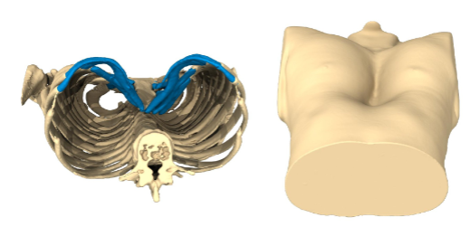
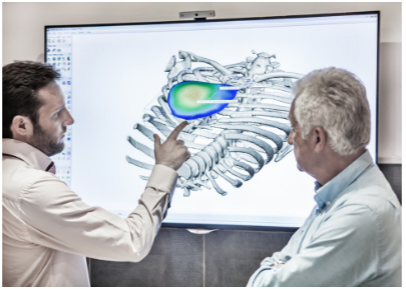
The implant is then surgically inserted inside the chest between the rib cage and the pectoralis muscles. An Artec Eva 3D scanner is then used to capture the new shape of the thorax. The pre-operation and post-operation scans are aligned, and distance map comparisons are performed in the Artec Studio scanning and processing software. This allows the medical team to see where and how body tissues are projected and enables them to understand how the soft tissues on top of the implants are repositioning themselves according to the standard anatomical structure of the rib cage.
Before they turned to 3D scanning, Moreno and his team would use the much more time-consuming and difficult process of plaster casting or CT scans to capture the patient’s outer anatomy. CT scans can only be performed while the patient is lying down, resulting in the soft tissues moving and creating inaccurate scans. A lot changed when AnatomikModeling brought in an Artec MH 3D and an Artec Eva. The company later acquired an Artec Spider to capture the heads and faces of patients in high resolution. While CT scans are still required to obtain data on patients’ internal anatomy, they are exposed to less radiation and their outer anatomical data can be taken in a standing position, achieving precise results.
“Using the Artec EVA and Spider VS classic plaster molding saves a lot of time and is much more reliable,” said Moreno. “Thus, it is much easier for the patient, compared to when plaster is used for molding. Also, having the 3D models of the patient’s anatomy allows you to take measurements that would be impossible to attain using the plaster molds.”
Discuss this and other 3D printing topics at 3DPrintBoard.com or share your thoughts below.
Subscribe to Our Email Newsletter
Stay up-to-date on all the latest news from the 3D printing industry and receive information and offers from third party vendors.
Print Services
Upload your 3D Models and get them printed quickly and efficiently.


You May Also Like
Johns Hopkins University Researchers Develop HyFAM Technology
Two scientists from Johns Hopkins University, Nathan C. Brown and Jochen Mueller, have developed a hybrid manufacturing technology they call HyFam, or Hybrid Formative Additive Manufacturing. Their work on this technology...
3D Printing G-Code Gets an Upgrade: T-Code
Good old G-Code still manages many 3D printers, great and small. Just like the STL, it’s a standard that enables collaboration while also holding the additive manufacturing (AM) industry back....
AM Rewind: The Biggest News and Trends of 2024
After a sluggish 2023, driven by persistent inflation and geopolitical tensions, 2024 has seen some recovery. Economic growth climbed from about 2.8 percent in 2023 to a modest 3.2 percent...
Metal Wire 3D Printer OEM ValCUN Announces Plans for 2025 Expansion
ValCUN, a Belgian original equipment manufacturer (OEM) of wire-based metal additive manufacturing (AM) hardware, has announced that the company has entered the next phase of its growth trajectory, making key...