

Subjecting 3D Printed Medical Ti6Al4V Implants to Laser Peening Could Increase Wear Resistance


When it comes to manufacturing biomedical implants, whether they’re 3D printed or not, titanium alloys are often used, and one of the most common is medical Ti6Al4V, thanks to its biocompatibility, mechanical properties, and excellent corrosion resistance. However, its use is not widespread due to its poor wear resistance. Additionally, the simultaneous action of both corrosion and wear could result in increased degradation of implants made with this alloy, as well as generate high wear debris, which limits stability. So it’s important to improve the alloy’s wear resistance in corrosive body fluid, in order to extend the service life of 3D printed implants made with medical Ti6Al4V.
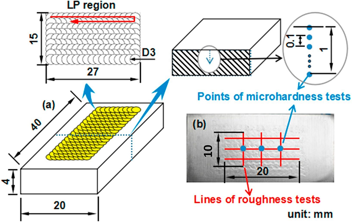
(a) A schematic view of LP path and (b) Detection program.
Many researchers are using surface modification techniques, like thermal oxidation and plasma surface alloying, to beef up the wear resistance. But the thermal effects of these methods can cause tensile residual stress in treated materials’ surface layer, which can then decrease fatigue strength.
![]() That’s why a group of researchers from Jiangsu University are turning to laser peening (LP), a laser surface modification technology, instead. They recently published a paper on their efforts, titled “Effect of laser peening on friction and wear behavior of medical Ti6Al4V alloy.”
That’s why a group of researchers from Jiangsu University are turning to laser peening (LP), a laser surface modification technology, instead. They recently published a paper on their efforts, titled “Effect of laser peening on friction and wear behavior of medical Ti6Al4V alloy.”
The abstract reads, “The aim of this study was to investigate the effects of laser peening (LP) on the friction and wear properties of medical Ti6Al4V alloy. Wear tests were conducted on the samples before and after LP with a ball-on-disk wear tester in Hank’s solution. The surface roughness and micro-hardness of the differently treated samples were measured. The friction and wear properties of the samples were evaluated by comparing their coefficient of friction (COF) and wear mass loss. The morphologies of wear scar and the wear mechanism were characterized by optical microscope (OM), scanning electron microscope (SEM) and energy dispersive spectrometer (EDS). The results showed that the surface micro-hardness of the specimen subjected to LP was significantly increased by 25.7% and the corresponding depth of the hardened layer was more than 0.3 mm. The average COF and wear mass loss were significantly reduced to 50% and 29.2%, respectively, as a result of the increasing laser energy and impact times. After LP, the wear mechanism turned from the severe oxidative wear to a slight adhesive wear and abrasive wear. Experimental results proved that LP treatment could effectively improve the friction and wear properties of the medical Ti6Al4V alloy.”
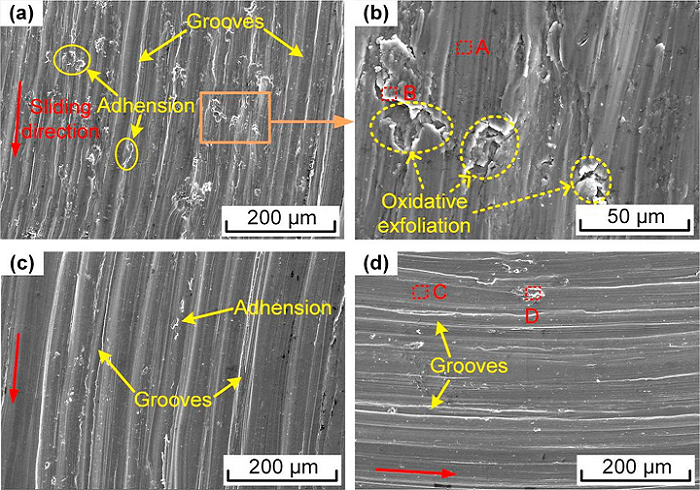
SEM micrographs of worn surface of untreated and LP treated specimens. (a) and (b) Specimen 0 (untreated); (c) Specimen 1 (1 impact-4 J-LP); (d) Specimen 6 (2 impacts-8 J-LP).
Laser peening uses severe plastic deformation, induced by plasma shock wave, to modify a material’s mechanical and microstructural properties, and other studies have shown that the method can refine the grain and improve microhardness of a material’s surface layer. When used with the proper treating parameters, LP can also improve the corrosion resistance and fatigue behaviors of metals and alloys – good news for 3D printed implants. But there hasn’t been a lot of research conducted on the wear and friction properties of medical Ti6Al4V alloy subjected to LP…until now, anyway.
“This paper aims to investigate the effects of LP on wear and friction behavior of medical Ti6Al4V alloy with different laser peening parameters in Hank’s balanced salt solution (HBSS),” the researchers explained. “Surface roughness and micro-hardness of the samples were measured, while their friction coefficient and wear mass loss were also investigated. Surface morphologies of wear scar on the sample were observed by a 3D optical microscope, and the wear mechanisms of the specimens before and after LP in simulated human body fluids were discussed based on SEM images and EDS data.”
The team used a commercially available medical Ti6Al4V alloy for the experiment, and cut samples into 40 x 20 x 4 mm rectangular shapes, with 4 mm thickness.
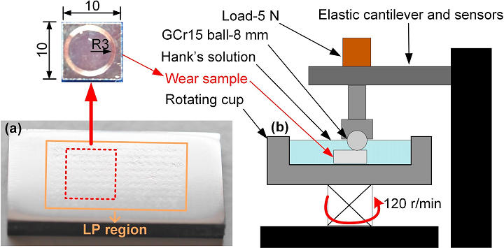
(a) Samples used in the wear test; (b) Schematic illustration of the wear test system.
Untreated and LP-treated Ti6Al4V specimens were tested for surface roughness and micro-hardness, and sliding wear tests were conducted at Nanjing University of Aerospace and Astronautics in China.
“During the test, the sample was immersed in Hank’s solution at room temperature and fixed in the rotating cup,” the researchers explained.
“The rotating cup was used to rotate the samples at a speed of 120 r/min for 30 min with wear radius of 3 mm. GCr15 ball (750 HV, Ra < 0.1 μm) of 8 mm diameter was used as the counter material. The test load was 5 N throughout the process which is closer to the condition of biomedical implant application. In all of the tests the COF were monitored against time directly through the elastic cantilever and sensors of this system.”

3D morphology (a–c) and section profile (d) of wear scar: (a) Specimen 0; (b) Specimen 1; (c) Specimen 6.
The samples were cleaned after the tests, and had their wear mass loss measured. Additionally, a 3D optical microscope was used to observe the 3D morphology of the wear scars, and SEM, combined with an energy dispersive spectrometer, observed their worn surfaces.
The team concluded that, when compared to the untreated sample, the surface micro-hardness value of the LP-treated Ti6Al4V sample increased by 25.7%, showing that LP was able to strengthen its surface. Additionally, the results of the experiment showed that after being treated with LP, the specimen did have better wear resistance than the untreated one; laser energy and impact time also helped improve the wear resistance of the LP specimen.
-
“After LP treatment, the wear mechanism of the Ti6Al4V specimen changed from severe oxidative wear into slight adhesive wear and abrasive wear,” the researchers wrote. LP process could contribute to the inhibition of toxic elements that are released in the human body when Ti6Al4V alloy implants are utilized.”
Co-authors of the paper are Jianzhong Zhou, Yunjie Sun, Shu Huang, Jie Sheng, Jing Li, and Emmanuel Agyenim-Boateng.
Discuss this and other 3D printing topics at 3DPrintBoard.com or share your thoughts below.
Subscribe to Our Email Newsletter
Stay up-to-date on all the latest news from the 3D printing industry and receive information and offers from third party vendors.
Print Services
Upload your 3D Models and get them printed quickly and efficiently.


You May Also Like
Aires Tide Designed with AI, Supercomputers, and 3D Printing
The Department of Energy‘s National Nuclear Security Administration (DOE/NNSA) is part of the US government that manages the US nuclear stockpile, helping to upgrade, improve, and maintain nuclear weapons, and...
3D Printing News Briefs, June 27, 2026: Nanoscale 3D Printing, Defense Readiness, & More
We’re starting with a story about a grant for advanced nanoscale 3D printing in this weekend’s 3D Printing News Briefs, and then on to metal additive manufacturing (AM) for defense...
US Army Awards Continuous Composites 3D Printed Missile Component Contract
Despite the very loud, indignant claims from American defense officials that the US hasn’t depleted a significant portion of its munitions stockpiles, the US has depleted a significant portion of...
Why Qualification Is Becoming the Next Frontier for AM in Energy
The energy industry doesn’t have much room for failure. Components used in power generation often operate under extreme temperatures and pressures, sometimes for decades at a time. That’s one reason...


