3D Printing Spotlight On: Vanessa Campanacho, PhD, Biological Anthropologist

 Vanessa Campanacho is a well-educated woman with a love for bones; in fact, she has attained educational degrees and is able to spend her days doing research some of us could only dream of—while many others don’t quite understand what a biological anthropologist does at all. I had to inquire further for a full understanding, as well as satisfying my curiosity about how she came to be so interested in the study of bones and along with it, skilled at death estimation in humans.
Campanacho received her Bachelor’s degree in 2008 from the New University of Lisbon, and then in 2010 her Master’s degree in Evolution and Human Biology from the University of Coimbra. In 2016, she received a PhD from the University of Sheffield, and now works as associated staff for teaching and research at the University of Sheffield, as well as a research associate in CIAS – Research Centre for Anthropology and Health and CEF – Centre for Functional Ecology – Group of Paleoecology and Forensic Sciences, University of Coimbra. And recently, she took some time out to speak with us about her research as well as how she uses 3D scanning to further her studies as we continue to highlight the work of women in technology.
-

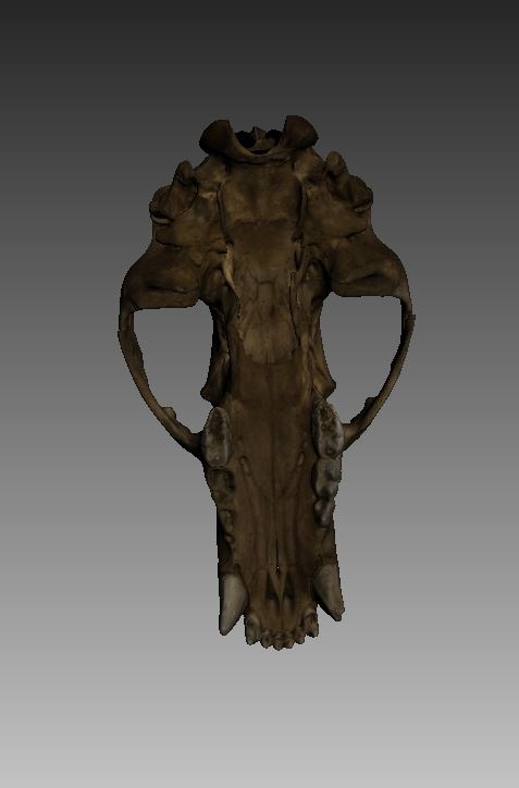
- 3D scans of a bear’s anatomy used for teaching purposes at the Department of Archaeology, University of Sheffield

“Initially, I was captivated by museology and anthropology of religion which led me to do a degree in anthropology at the New University of Lisbon, Portugal. However, after attending Professor Cláudia Sousa’s class, I became fascinated by biological anthropology, and couldn’t picture myself doing anything else.
With the support of Professor Sousa, I got the opportunity to become a volunteer at the National Museum of Natural History and Science in Lisbon. Under the guidance of Dr. Hugo Cardoso, I learned to distinguish each bone in the human skeleton and basic biological anthropological methods, and started my first steps in research. Additionally, I helped in the curation of the Lisbon identified skeletal collection by marking their collection number.
An identified skeletal collection is a skeletal assemblage whose biographical information is known, like sex, age, occupation and cause of death. Those collections are a valuable resource for biological anthropology research. The majority of methods were established and tested with an identified skeletal collection to be applied into unidentified skeletal remains from archaeological and forensic contexts.
Later on, I became a volunteer at an archaeological laboratory (CIPA) also during my degree. Under the guidance of Dr. David Gonçalves, I had the opportunity to excavate Iron Age cremations urns. I am very grateful for the experience I obtained during my undergraduate years.”
Do you find that many are not quite sure what it is you do?
“I would say the vast majority of people I meet initially are not quite sure what biological anthropology is. However, when I explain I study human bones which can be applied in archaeological and forensic remains, I see an increased interest and curiosity about my work. Successful television programs, such as Bones and CSI, usually tend to increase the interest of the greater public in this field. But I also found that the use of 3D scanning in my research also makes the greater public eager to learn more about it, not just how the technology works, but its research application.”

Dr. Campanacho showcasing the Artec Eva 3D scanner and the 3D modelling research done at the Department of Archaeology at the Faculty Showcase, at the Millennium gallery
How do you define your work to the general public?
“Working with the greater public is a rewarding experience, different from working in a laboratory, but equally gratifying. Being able to interact with the greater public allows me to talk directly about my research, including 3D scanning.
A recent experience was during the Faculty of Arts and Humanities showcase at the Millennium gallery. An event which brought the University of Sheffield closer to the community. I am grateful to have had the opportunity to represent the Glover laboratory for Digital Osteology from the Department of Archaeology. I got to showcase the Artec Eva scanner from Artec 3D by scanning hands of the public.
Artec Eva is a white LED structured light scanner which creates accurate three dimensional digital replicas of a bone with a resolution of up to 0.5 mm. The Glover laboratory also has Artec Spider, a blue LED structured light scanner with a resolution of 0.1 mm and ideal for smaller objects with more detail. However, both Artec scanners can be used on the same project. The vast majority of the public were not just attracted to see their hand on the screen, but were eager to know how the equipment worked and its application in biological anthropology. It is amazing to feel the enthusiasm of the greater public when learning about biological anthropology research and 3D scanning.
Additionally, I am the co-editor of Show Us Your Research!, an anthropological and archaeological publication for the great public. This free on-line publication allows the opportunity to create a link between the research produced and the greater public. Public engagement should always be part of a researcher’s work.”

The Artec Eva 3D scanner
What makes your research important now, and for posterity?
“I have been investigating which environmental factors affect skeletal ageing in adult individuals. After the skeleton stops growing and maturating, it starts suffering degenerative changes with age, such as the metamorphosis at the pelvic joints surface. Methods have been created to estimate the age an adult individual had at the time of death by looking at those degenerative changes. However, those methods have shown to not be accurate creating one of the biggest methodological problems in bioanthropology. The lack of accuracy is due to the variable degeneration rate between individuals and populations.
Even though the exact cause of the variability is largely unknown, researchers are now investigating what environmental and genetic factors are affecting the skeletal ageing. In my PhD thesis, I investigated the effect of body size on the degeneration at the pelvic joints with age in two skeletal samples, one from Portugal and another from the United States. Stature and body size was estimated by measuring the femur, one of the bones more correlated with these body size measures. Additionally, I used a structured light scanner which allowed computing the surface area of the joints which is not possible to be calculated with traditional anthropometric techniques.
The research gave more information about the ageing skeletal process, since body size affects age-related criteria at the pelvic joints. Investigating what factors are affecting the skeletal ageing process will help to improve the age at death estimation methods. It is imperative to estimate the age of an adult individual with accuracy in forensic context in order to help to identify a potential victim. For archaeological remains by estimating a reliable age at death allows to create reliable demographical and health profiles of past communities.”
 Do you work with many other women, as peers, in bioanthropology?
“Yes, I have been very lucky to work with amazing female researchers, but also to be supervised by remarkable female researchers at my Master’s and PhD. They have set great examples for other female researchers in my field—an example I now share with my students. At the moment, I am supervising female master students, and three of them will be using Artec 3D scanners in their dissertation projects. Their projects look at different topics within the biological anthropology field, as facial reconstruction, age estimation and sex diagnosis in immature ilia. It is a rewarding experience to be supervising while they mature as researchers.”
Was there a learning curve as you began using the Artec Spider and Eva 3D scanners to create models at the University of Sheffield?
“I have used a structured light scanner from FlexScan 3D on my PhD thesis, making me already familiar with common functions between scanners, such as mesh, alignment, fusion, smoothing and decimation. Although I already had experience in scanning, there was a learning phase when I began using the Artec Spider and Eva scanners to scan each type of bones in the human skeleton. The scanning procedure can imply some differences between different types of scanners. The FlexScan 3D scanner entails being fixed in a calibrated space while the bone is moved to capture its different surfaces. Eva and Spider Artec 3D scanners operate in a different way. While Artec 3D scanners can be handled and moved around the bone while capturing data, it can also be fixed in one position while the bone is moved to capture its different surfaces. After my learning phase with Artec 3D scanners, I wrote a guidelines document to scan bones which will be available on my website soon.”
Will you be using this technology in the classes you teach?
“I have incorporated scanning with Artec 3D scanners in my teaching before, allowing students to have a first contact with this technology which they may later on use on their dissertations research.”

A figure from 3D scanning guidelines Dr. Campanacho developed
Do you find that your colleagues are surprised you are using such technology, or are they using it as well more commonly now?
“The use of 3D scanning technology has becoming widely dispersed in biological anthropology and more departments are acquiring scanners to use in teaching and research. The Department of Archaeology, at the University of Sheffield, acquired Artec 3D scanners, with the aim to employ it in teaching, outreach and into the different fields of research that are being performed at the department. Artec 3D scanners allow scanning different sizes range for different applications within the department, but also to establish research connections with other universities.”
How do you see 3D technology changing the face of research, sharing, and archiving in bioanthropology?
“The use of scanning technology is not a novelty in biological anthropology, but its use has become widespread due to its increased accessibility associated with a higher mobility and resolution. In research, a new trend emerging is the automated analysis through 3D digital models to help perform the analysis without the subjective analysis of the researcher.
This type of analysis aims to diminish the observer error which may be present in some techniques. I intend to establish a sex diagnosis method by quantifying the volume of cranial features from 3D digital models. Sex diagnosis of non-identified skeletal remains may be performed by directly observing cranial morphological traits, with females’ individuals being more gracile than males. However, some crania show a mix of male and female features making it difficult to diagnose their sex. In light of that, the quantification of the volume in several cranial features may help to diagnose sex from 3D digital models instead of the direct observation on the crania.
Another trend regards the sharing of 3D digital models of human bones among researchers and the greater public. 3D digital models can potentially be shared across the globe facilitating research without having the costs associated with traveling. Furthermore, it will enhance research and increase interactions between investigators. Researchers are still discussing the best practice to sharing 3D digital models though. Questions have arisen regarding access, copyright and ethical issues. It can be argued that it is important to make the models accessible for the research community and the greater public, to share and produce knowledge. However, it is equally vital to know the aim the user will have in the manipulation of human bone replicas. Institutions that curate human remains are responsible for their integrity and protection, and thus need to ensure that the same is enforced into the 3D digital and printed replicas.”
-

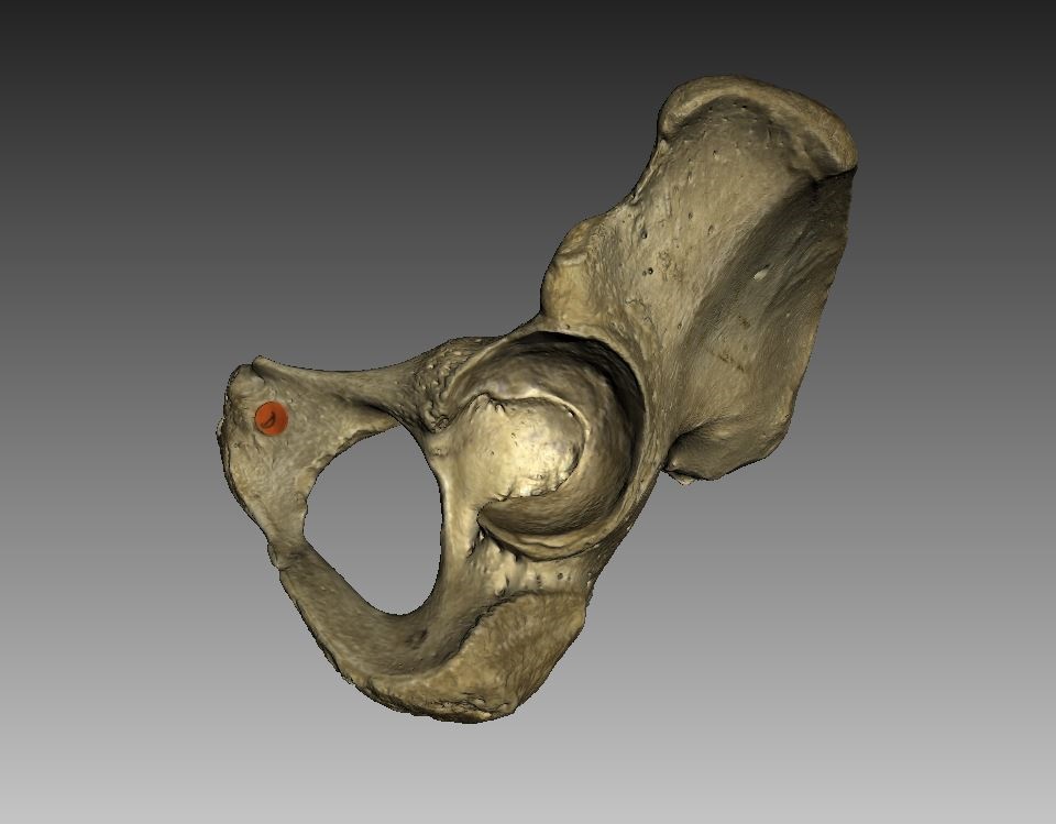
- Human left pelvic bone
-
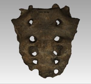
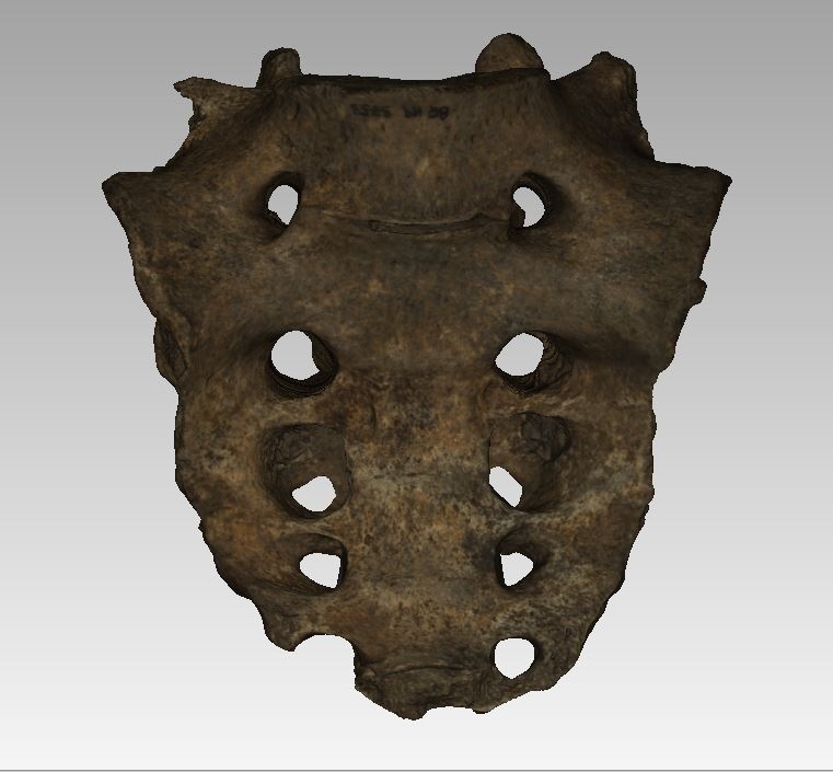
- Sacrum
The work Dr. Campanacho is engaged in has broad reach, and great potential for the study of humanity—helped along through 3D scanning and other high-tech means. Work being done at the University of Sheffield often employs 3D technologies from talented researchers. We’re sure to see technology continue to play a great role in anthropological and other research, and look forward to following Dr. Campanacho’s future achievements. Share your thoughts in the Vanessa Campanacho forum at 3DPB.com.
If you are interested in sharing your story, or know a woman we should get in touch with for this new series, please reach out any time. Send us an email or connect on Twitter. We’re looking forward to sharing more stories about women in 3D printing. Find all the features in this series here.
[Images: Dr. Vanessa Campanacho]
Subscribe to Our Email Newsletter
Stay up-to-date on all the latest news from the 3D printing industry and receive information and offers from third party vendors.
Print Services
Upload your 3D Models and get them printed quickly and efficiently.


You May Also Like
The Market and Industry Potential of Multi-Material 3D and 4D Printing in Additive Electronics
Additive manufacturing leverages computer-based software to create components for products by depositing either dielectric or conductive materials, layer by layer, into different geometric shapes. Since its birth in the 1980s,...
3DPOD 262: Bio-inspired Design for AM with Dhruv Bhate, Arizona State University
Dhruv Bhate is an associate professor at Arizona State University. There, he looks at structures, materials, and design. Previously, he worked at PADT as well as in the semiconductor and...
3DPOD 261: Tooling and Cooling for AM with Jason Murphy, NXC MFG
Jason Murphy´s NXC MFG (Next Chapter Manufacturing) is not a generalist service; instead, the company specializes in making tooling. Using LPBF and binder jet, the company produces some of the...
3DPOD 260: John Hart on VulcanForms, MIT, Desktop Metal and More
John Hart is a Professor at MIT; he´s also the director of the Laboratory for Manufacturing and Productivity as well as the director of the Center for Advanced Production Technologies....