
New 3D Bioprinting Method Could Lead to More Effective Treatment for Osteoarthritis

 Everyone knows that arthritis is an all-too-common – and painful – condition, but the most common form is osteoarthritis, also known as degenerative arthritis or degenerative joint disease. The condition is a result of the breakdown of cartilage and is, needless to say, extremely painful and debilitating. There are several methods of treating the condition, but their effectiveness varies from case to case, and no cure exists – it’s just symptom management once the disease has been diagnosed.
Like with so many other diseases, however, treatment for osteoarthritis may become much more effective before long – thanks, of course, to 3D printing. Researchers at Penn State University are developing a new method of bioprinting that could eventually be used to create cartilage patches for worn out joints – a potential cure for osteoarthritis.
“Our goal is to create tissue that can be used to replace large amounts of worn out tissue or design patches,” said Ibrahim T. Ozbolat, associate professor of engineering science and mechanics at Penn State. “Those who have osteoarthritis in their joints suffer a lot. We need a new alternative treatment for this.”
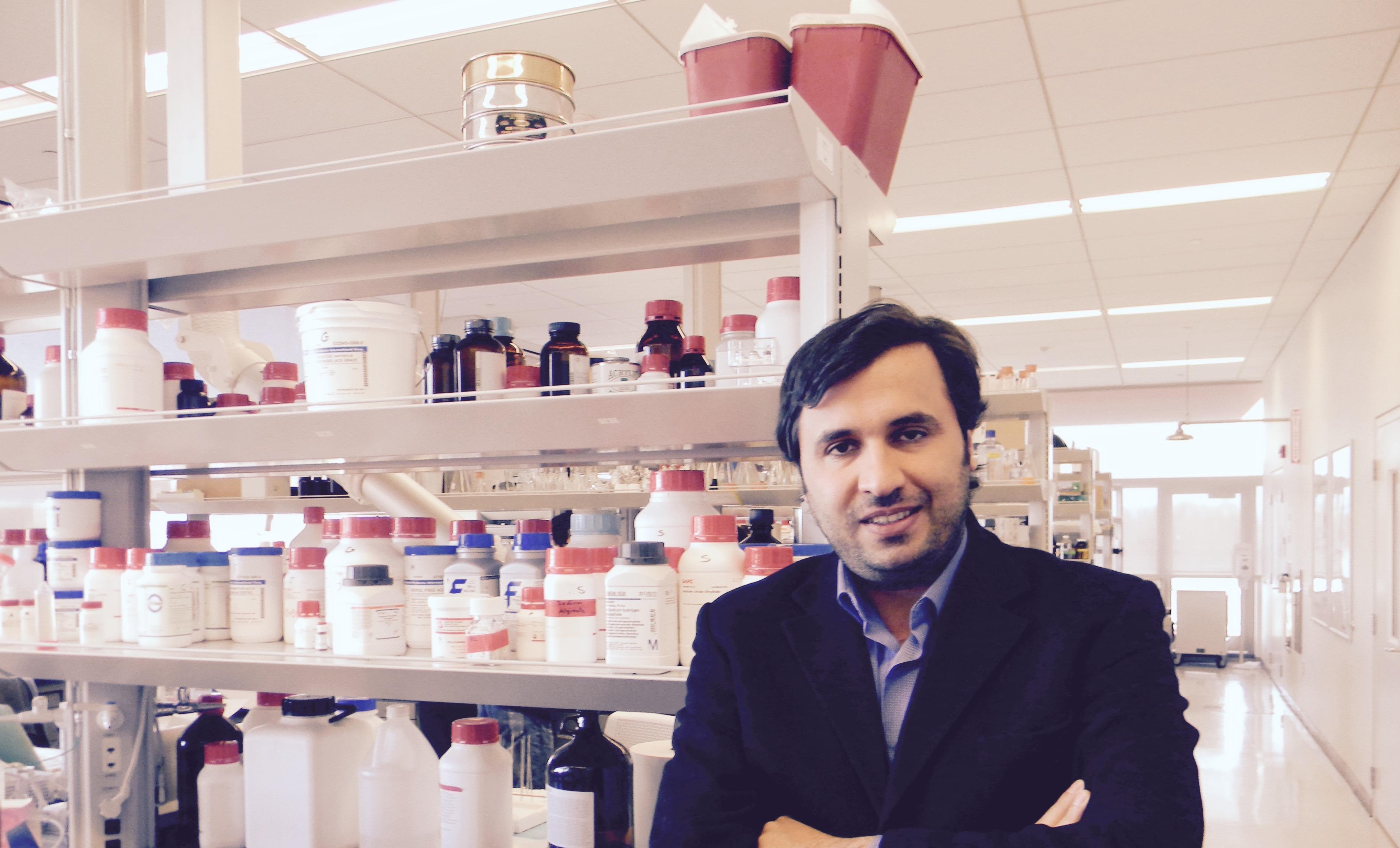
Ozbolat in his lab.
The difficulty in treating osteoarthritis lies in the fact that cartilage cannot repair itself – once it’s gone, it’s gone. It also happens to be one of the easier tissue forms to reproduce via 3D printing, however – it has no blood vessels, and consists of only one type of cell, making it an ideal place to start when experimenting with new methods of bioprinting.
No form of bioprinting is simple, though, and the Penn State researchers are working to develop a method that circumvents one of the more common frustrations encountered when trying to 3D print human tissue. Most methods involve the creation of a scaffold by embedding cells in a hydrogel, which then – ideally – grows into living tissue. That’s not always what happens, though, unfortunately.
“Hydrogels don’t allow cells to grow as normal,” said Ozbolat. “The hydrogel confines the cells and doesn’t allow them to communicate as they do in native tissues.”
Ozbolat and his team, therefore, decided to avoid hydrogels altogether and take a different approach. They created a tiny tube, about three- to five-hundredths of an inch in diameter, made from an algae extract called alginate. Cartilage cells are injected into the tube and allowed to grow and adhere to each other for about a week, at which point the alginate tube is removed – easy to do, as the cells do not stick to the algae material. The team is then left with a strand of cartilage that acts as, essentially, living filament.
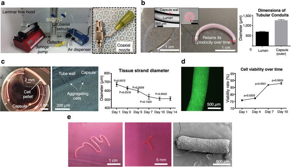
The researchers designed a special prototype printer nozzle into which the cartilage strands are fed and then extruded, layer by layer. After only half an hour or so, the strands adhere to each other enough that the whole printed structure can be moved to a petri dish, where it is provided nutrients and allowed to further grow and fuse into a piece of cartilage. So far, the cartilage produced by the Penn State team has been inferior to natural cartilage, but better than cartilage that has been produced using hydrogel – plus, the process allows for a lot more flexibility.
“We can manufacture the strands in any length we want,” said Ozbolat. “Because there is no scaffolding, the process of printing the cartilage is scalable, so the patches can be made bigger as well. We can mimic real articular cartilage by printing strands vertically and then horizontally to mimic the natural architecture.”
 The cartilage printed in Ozbolat’s lab is similar to cow cartilage; if the process ends up being used for human procedures, cells from each individual patient would likely be used as “seed” material to prevent the risk of rejection. Ozbolat also believes that as natural cartilage forms with pressure from the joints, the properties of 3D printed cartilage will improve when pressure is applied.
Ozbolat and his team – including Yin Yu, Kazim K. Moncal, Jianqiang Li, Weijie Peng, Iris Rivero, and James A. Martin – published their research in a just-released paper in Scientific Reports. Like any new medical technology, the method has a long way to go before it can be used in any kind of human trials, but it looks promising, and has the potential to eliminate a great deal of pain for many, many people in the future. Discuss this new method further in the BioPrinting for New Joints forum over at 3DPB.com.
Subscribe to Our Email Newsletter
Stay up-to-date on all the latest news from the 3D printing industry and receive information and offers from third party vendors.
Print Services
Upload your 3D Models and get them printed quickly and efficiently.


You May Also Like
Metal Powder Supplier Elementum 3D Added to $46B Air Force Contract
Elementum 3D, a Colorado-based developer and supplier of metal powders used in additive manufacturing (AM), announced that the company has been added to the vendors list in the fourth on-ramp...
Ursa Major Lands $28.6M AFRL Deal for 3D Printed Draper Engine Flight Demo
The US Air Force Research Laboratory’s (AFRL’s) Rocket Propulsion Division at Edwards Air Force Base has awarded a $28.6 million contract to Ursa Major for follow-on work related to the...
3D Printing Financials: Rocket Lab’s Record-Breaking Year and Over 20 Launches Coming in 2025
Rocket Lab (Nasdaq: RKLB) closed 2024 with its best year yet. The company launched more rockets, signed more contracts, and expanded deeper into spacecraft and satellite production than ever before....
US Air Force Taps Beehive to Study 3D Printed Jet Engines
Propulsion 3D printing firm Beehive Industries secured a contract from the U.S. Air Force Life Cycle Management Center through SOSSEC. SOSSEC is a company that manages Other Transactions Authority (OTA)...