Surgeons Use HoloLens and Metal 3D Printing to Implant Eye Socket

Both augmented reality (AR) and 3D printing have been used by surgeons over the past decade, as teaching tools for medical schools and as patient-specific 3D maps for performing surgeries. These 3D models are processed from a patient’s anatomy using dense data captured from computed tomography (CT) scans. But rarely are 3D printing and AR used in concert by surgeons to improve the accuracy, quality and efficiency of surgeries.
Yet a team of doctors at Galilee Medical Center for Oral and Maxillofacial Surgery recently performed a repair of a fracture in the floor of an eye socket using augmented reality and 3D printing. A 31-year-old patient underwent surgery led by Prof. Samer Srouji of Galilee Medical Center’s Center for Oral and Maxillofacial Surgery. The patient fractured the left eye socket of his face leaving him impaired with double vision and facial disfiguration.
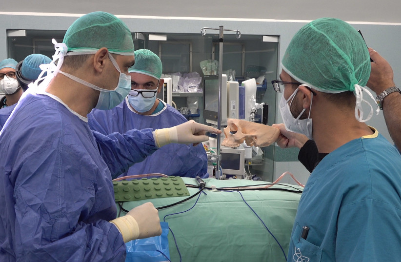
Image courtesy of the Jerusalem Post.
In a statement to the Jerusalem Post, Prof. Srouji said, “The innovative technology utilizing a 3D printer and augmented reality resulted in both a particularly accurate execution of the operation, and a significant reduction in time.”
The patient received a 3D printed titanium plate based on a 3D model built from scanned CT data of the injured left socket area. The scan was an inverted version of the patient’s healthy right socket area, projected onto the injured left socket area with 3D software.
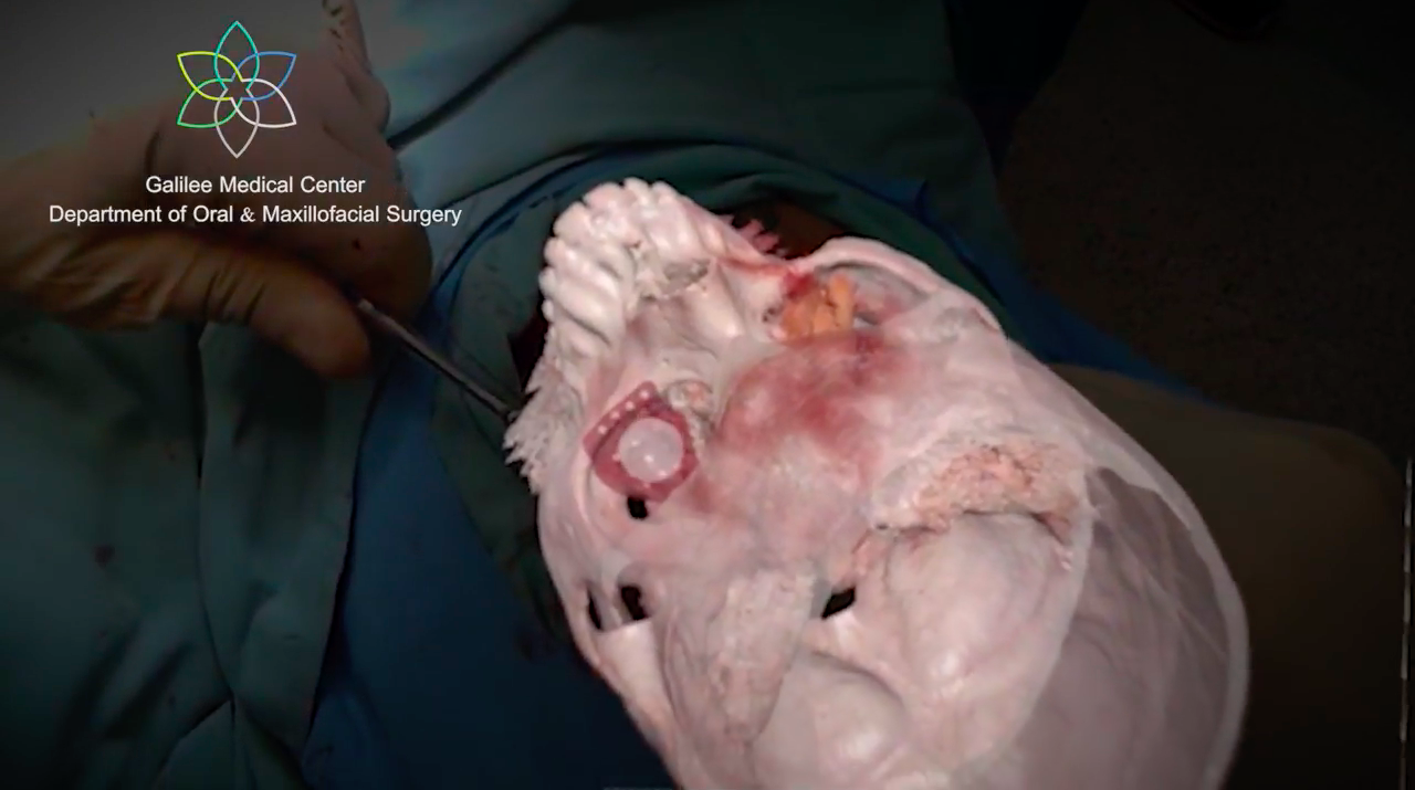
A screenshot of the operating procedure from the perspective of the HoloLens. Image courtesy of the Jerusalem Post.
To position the 3D printed titanium plate correctly, one doctor used Microsoft HoloLens mixed reality glasses to overlay the 3D model of the titanium implant into position under the patient’s left eye socket. Overlaying the 3D models of the patient’s skull and the 3D model of the 3D printed titanium plate on the patient’s actual facial anatomy, doctors at Galilee Medical Center accurately positioned the implant into the existing bone structure. A brief video of the procedure below shows the utility of applying AR in the operating room.
After just 90 minutes, the surgery was over and, within a few days, the patient was released from the hospital. Following recovery of several days, the patient was released home. The patient’s follow-up CT scans revealed that the 3D printed titanium plate implantation was successfully placed without any complications.
Subscribe to Our Email Newsletter
Stay up-to-date on all the latest news from the 3D printing industry and receive information and offers from third party vendors.
Print Services
Upload your 3D Models and get them printed quickly and efficiently.


You May Also Like
AM Asia Watch: China’s HeyGears Lands $44M to Expand Beyond Dental 3D Printing
Chinese 3D printing company HeyGears raised more than 300 million Yuan (roughly $44 million) in a new Series C funding round as it looks to expand beyond its industrial and...
The University of Utrecht: “3D Printing Could Change Who Gets to Become a Manufacturing Power”
For decades, manufacturing has mostly been controlled by countries with huge factories, lower labor costs, and industrial systems that took years, sometimes decades, to build. But Utrecht University human geographers...
3D Printing News Briefs, May 28, 2026: Continuous Fiber Reinforcement, Bioprinted Trachea, & More
In today’s 3D Printing News Briefs, America Makes announced the winners of its JAQS-SQ Project Call. Axtra3D is partnering with Keystone Industries to expand its dental material ecosystem, while BigRep...
Asia AM Watch: China’s SHINING 3D Restarts IPO Review Process
SHINING 3D is moving forward again with its plans to go public in China, after restarting its Beijing Stock Exchange (BSE) initial public offering (IPO) review process and filing updated...